
Figures & data
Figure 1 Preparation of the snail-tipped exoplant. (A) A malleable titanium plate (0.5mm thick, 20 holes, titanium adaption plate, MatrixMIDFACE™ Plate, Depuy Synthes, West Chester, PA, USA) was used to strengthen the exoplant. (B) After tunneling the silicone sponge (506 silicone sponge, MIRA, USA) with a 20-gauge MVR blade and the titanium plate was inserted into the tunnel. (C) When the titanium plate was reached to the end of the silicone sponge, the tip was rolled to a snail shell shape.
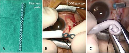
Figure 2 Location of the exoplant. A schematic drawing shows the location of the exoplant in a left eye. The exoplant is inserted superotemporally to reach the macular area.
Abbreviations: IR, inferior rectus muscle; LR, lateral rectus muscle; MB, macular buckle; MR, medial rectus muscle; SR, superior rectus muscle.
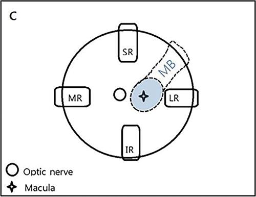
Figure 3 Stepwise approach of the surgical procedure. (A) After 120° superotemporal conjunctival peritomy, bridle suture was passed under the superior rectus and the lateral rectus muscle. (B) 25-gauge PPV and ILM peeling using MaQaid® for chromovitrectomy was performed. (C) Rotating the eyeball inferonasally, the prepared exoplant was inserted superotemporally to the posterior of the globe to reach the macula. (D) The buckle height and location was adjusted under direct visualization through the vitrectomy lens. (E) The long arm of the exoplant was fixed with a mattress suture posterior to the muscle insertion sites. (F) The buckle height and location was checked once again. (G) A second mattress suture was fixed posterior to the first mattress suture. (H) The remnant length of the exoplant was trimmed using a plate cutter. (I) The titanium plate was cut more deeply into the silicone sponge to hide its end into the sponge. (J) Fluid air exchange and subretinal fluid drainage was done. (K) Gas or silicone oil tamponade was performed. (L) Finally, the conjunctiva was approximated with 8–0 Vicryl suture.
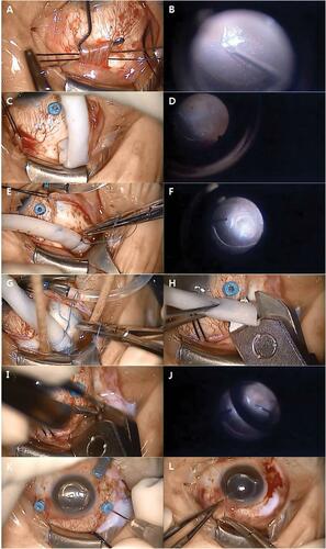
Table 1 Demographic And Other Baseline Characteristics Of Patients Before And After Surgery
Figure 4 Pre- and postoperative OCT image of a 71-year-old woman (case 1). (A) MHRD with ERM is seen on preoperative OCT. The excavation of the posterior sclera was too deep to be captured simultaneously. (B) 30 months postoperative OCT shows reattachment of the retina with persistent MH. The buckle effect is well noticed on the OCT.
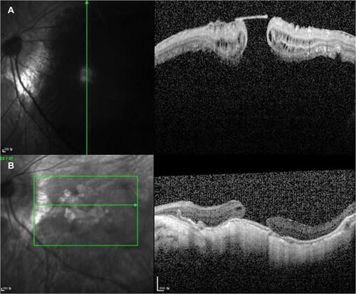
Figure 5 Pre- and postoperative OCT image of a 61-year-old woman (case 4). (A) Preoperative OCT shows a high myopic eye with MHRD and PS. (B) 24 months postoperative OCT shows flattening of the posterior sclera with MH closure and retinal reattachment.
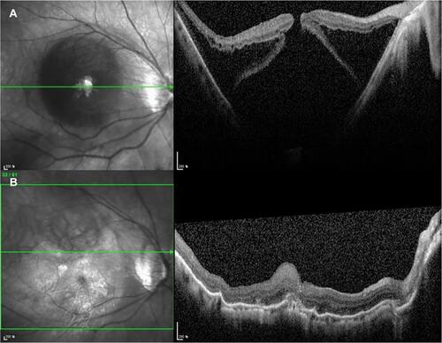
Figure 6 Pre- and postoperative OCT image of a 55-year-old woman (case 7). (A) Preoperative OCT shows foveal detachment and retinoschisis with MH. (B) 11 months after the surgery, MH closure was achieved with decreased bulging of the posterior scleral wall was seen on postoperative OCT.
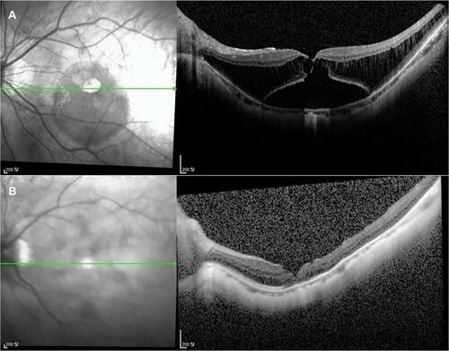
Figure 7 Pre- and postoperative B-scan USG images. (A) Preoperative B-scan USG image shows PS with retinal detachment (case 3). (B) Postoperative B-scan USG image shows correction of PS with indentation of the posterior sclera due to macular buckle effect (case 3). (C) Preoperative B-scan USG image shows PS with retinal detachment (case 4). (D) Postoperative B-scan USG image shows correction of PS with indentation of the posterior sclera due to macular buckle effect (case 4).
