Figures & data
Figure 1 (A) Air escape from the perforation. (B) Air escape from the perforation displacing the previously excised stromal tissue which was used for external tamponade, the arrows show the boundaries of the perforation (Intraoperative picture).
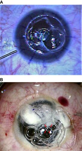
Figure 2 (A) Stable anterior chamber filled with air after suturing the previously excised stromal tissue the circle delineate the sutured whole cap of the previously excised stromal tissue. (B) Stromal dissection using Barraquer spatula while the anterior chamber is filled with air (intraoperative picture).
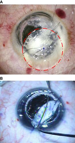
Figure 3 (A) Completion of stromal dissection near the pre-descemetic plane at the Descemet’s membrane tear with a scissor. (B and C) Applying the graft after completion of stromal dissection to a pre-descemetic plane (Intraoperative picture).
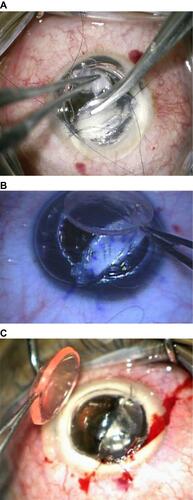
Figure 4 (A–C) The corneal graft is secured in place with a few interrupted sutures while pulling the stromal patch through the edge of the trephine. (D) another case. (Intraoperative picture) Pulling stromal patch out through the trephine edge (Intraoperative picture).
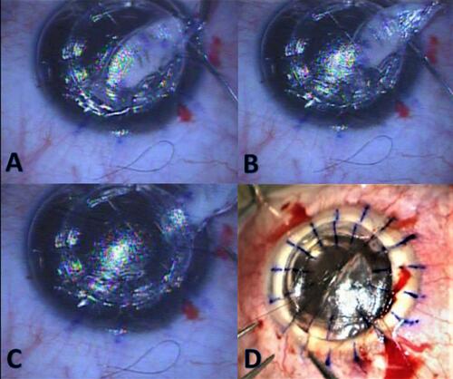
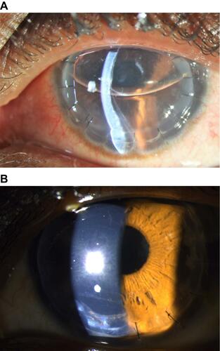
Figure 5 (A) Slit lamp photo three days after surgery of one case with large inferior Descemet’s membrane tear showing a detachment. (B) Same eye 18 months after surgery showing the large Descemet’s membrane break with clear stromal tissue and interface.