Figures & data
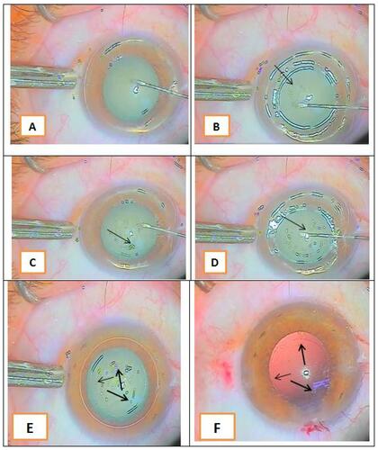
Figure 1 (A) Starting capsulorhexis; (B) multiple light reflection rings at different tissue levels (arrow shows the capsulorhexis edge); (C) progression of capsulorhexis guided by light reflection at the torn capsular edge (arrow shows the capsulorhexis edge); (D) closing of capsulorhexis (arrow shows the capsulorhexis margin to the end of the circle); (E) arrows showing the completed circular capsulorhexis; and (F) arrows showing the complete capsulorhexis against a red reflection after phacoemulsification.