Figures & data
Figure 1 A photograph of Gomphocarpus physocarpus fruits. Inflated fruits of Gomphocarpus physocarpus have a characteristic balloon-like shape. The fruit is approximately 5 cm long and wide and it contains numerous small black seeds.
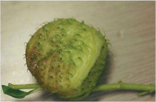
Table 1 Patients’ Basic Characteristics And Clinical Findings On The First Visit
Figure 2 The changes in central corneal thickness of the patients. Increased central corneal thickness was observed at the first visit and it gradually decreased to approximately less than 600 μm 2 weeks after the injury.
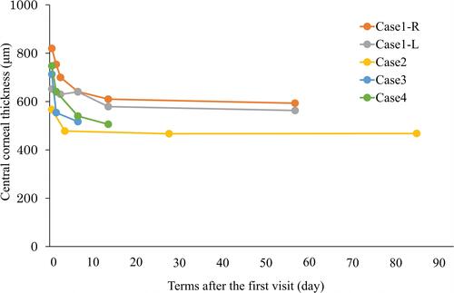
Figure 3 The changes in corneal endothelial cell density of the patients. Corneal endothelial cell density was higher than 2000 cells/mm2 in all patients at the first visit and during the observation period.
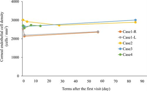
Figure 4 Clinical findings of the anterior segment at the first visit for case 1. (A, C) Photograph of the anterior segment of the right (A) and left (C) eyes. Folds of Descemet’s membrane were observed. (B, D) Photograph of the cornea of the right (B) and left (D) eyes after the use of fluorescein. Superficial punctual keratitis was bilaterally observed but corneal epithelial defect was not observed.
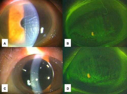
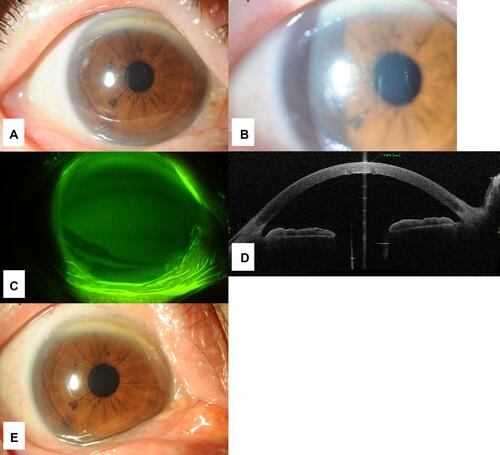
Figure 5 Clinical findings of the anterior segment at the first visit for case 4. (A) Photograph of the anterior segment of the right eye. Hyperemia was not observed. (B) Photograph of slit-lamp examination of the right eye. Folds of Descemet’s membrane were observed. (C) Photograph of the cornea after the use of fluorescein. Corneal epithelial defect was not observed. (D) Optical coherence tomography image of the anterior segment. Paracentral corneal thickness was 625 μm and corneal edema was observed. (E) Photograph of the anterior segment after the treatment. The folds of Descemet’s membrane disappeared.