Figures & data

Figure 1 (A–C) Corneal channel dissection parameters programmed into the Alcon/WaveLight FS200 femtosecond laser. (D) Anterior segment OCT illustrates the – as planned – position within the host cornea, of the implanted xenograft intracorneal ring segments.
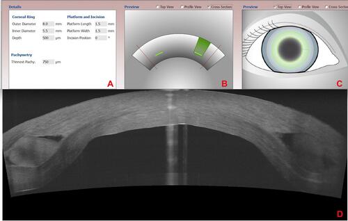
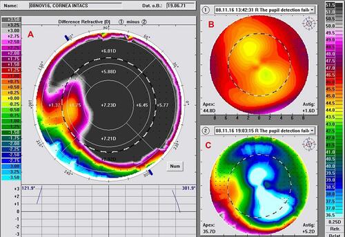
Figure 2 These images illustrate the Placido-disc topographic changes documented in this ex-vivo procedure. Specifically: (A) reflects the difference flattering achieved (an impressive 7 Diopters) with the combined ICRS xenograft and CXL from (B) pre-operative to (C) post-operative.