Figures & data
Figure 1 Fundus photograph of the left eye of a 33-year-old female with biallelic RPE65 gene mutation demonstrating diffuse pigmentary changes, vascular attenuation, and temporal disc pallor.
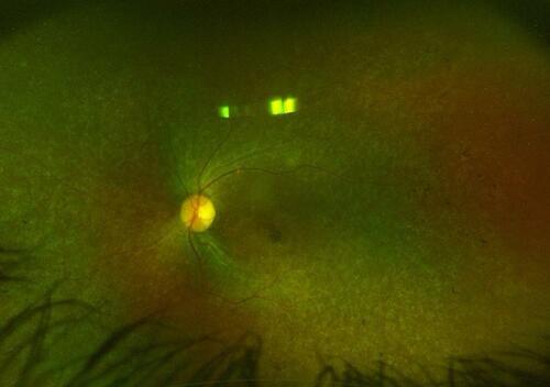
Figure 2 Optical coherence tomography images of the right (A) and left (B) macula of a 6-year-old boy with biallelic RPE65 gene mutation. Notice the diffuse loss of the ellipsoid zone except for a small subfoveal island in both eyes. Also note the marked atrophy of the outer nuclear layer (arrows) which slightly increases in thickness in the parafoveal and foveal regions.
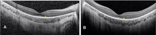
Table 1 Summary of the Major Human Clinical Trials Evaluating RPE65 Gene Therapy
Figure 3 Image illustrating the set-up of the injection apparatus. A 1-mL syringe containing the prepared voretigene neparvovec is connected to extension tubing and a 41-gauge subretinal injection cannula.
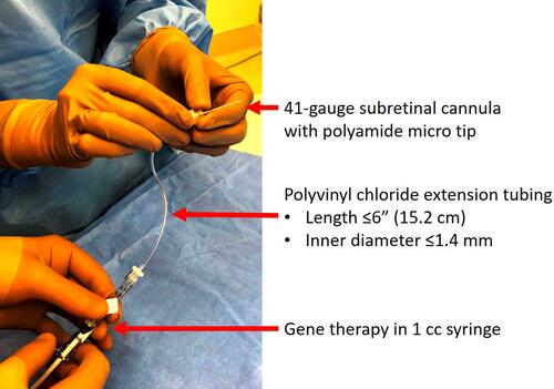
Figure 4 Intraoperative photograph illustrating a fovea-involving subretinal bleb created by injecting 0.3 mL of voretigene neparvovec.
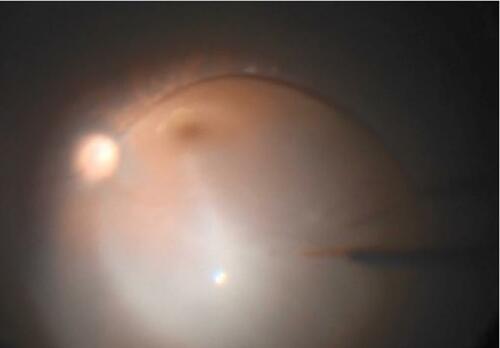
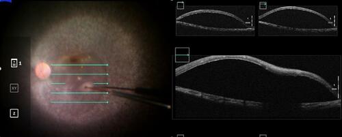
Figure 5 Intraoperative optical coherence tomography imaging can be used to guide the formation of a subretinal bleb of voretigene neparvovec and confirm its proper localization in the subretinal space.