Figures & data
Figure 1 Videokeratographic map of the right eye of a 32 yrs old male patient treated with PRK with the MEL-70 excimer laser (Carl Zeiss Meditec). Preoperative refraction was −4.50sph with a DCVA of 20/20. Eight months after PRK, his DCVA was 20/20 with −0.25sph. The treatment was well centered and effective.
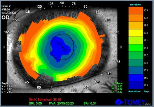
Figure 2 Right eye videokeratography maps of 35 yrs old female patient, originally treated with an unknown excimer laser using a standard PRK. Preoperative MRSE was −6.50 D with a DCVA of 20/20. Eight months after PRK, the refractive error was +2.00sph (DCVA 20/25), with a 1.2mm decentration of the photoablation.
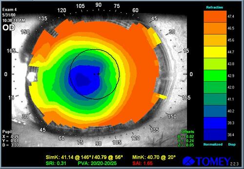
Figure 3 Right eye videokeratographic maps of a 41 yrs old female patient, before (A) and after (B) enhancement 2 years post-PRK using topographically guided customized excimer laser photoablation. After PRK her DCVA was 20/22 with −1.50sph=−1.50cyl (130°). Fifteen years after enhancement using topographically guided excimer laser photoablation with Corneal Interactive Programmed Topical Ablation platform (CIPTA, LIGI, Taranto, Italy) her DCVA was 20/20. The topographic values highlight the improved corneal profile obtained.
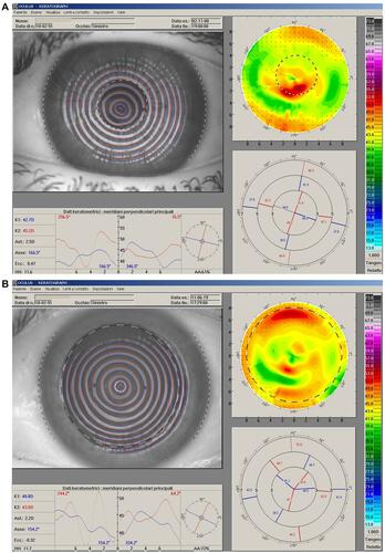
Figure 4 Right eye videokeratography maps of 64 yrs old female patient, before (A) and after (B) enhancement 3 years post-PRK using topographically guided customized excimer laser photoablation with CIPTA platform. The topographic values highlight the improved corneal profile obtained. Prior to transepithelial photoablation her DCVA (−2sph=−1 cyl (30°) was 20/25. After the procedure her DCVA was 20/20 with −0.50sph.
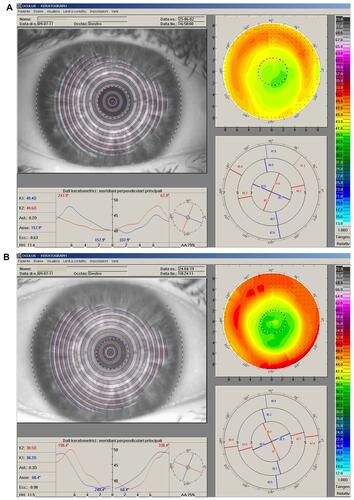
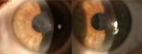
Figure 5 The figure shows slit lamp examination of a female patient 54 yrs old who had developed an evident haze 2 months after PRK. At that time her DCVA was 20/30 with −0.50sph=−3.50cyl (130°). After 8 months she underwent scraping of the stromal surface and application of mytomicin C. The images show left eye slit lamp photographs before (left) and 6 months after (right) the treatment. The corneal opacity is remarkably reduced, and her DCVA is now 20/25 with −0.50sph=−0.50cyl (150°).