Figures & data
Table 1 Patient Characteristics and Surgical Details
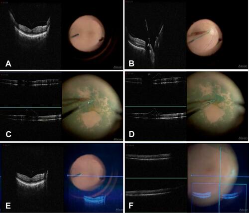
Figure 1 Screen grabs from a left eye live video demonstrating simultaneous same screen iOCT viewing with the Haag Streit (gen 2) iOCT side by side with the live surgical field during a membrane peel in a patient with vitreomacular traction and ERM. (A) Vitreomacular traction at fovea, (B) Posterior hyaloid attached at optic nerve, C-D) Membrane peel, (E) Vitreomacular traction at fovea with OCT image injection, (F) Macula following membrane peel with OCT image injection.