Figures & data
Table 1 Changes in a-Wave, b-Wave, and 30 Hz Flicker in Different Time Points
Table 2 Changes in Scotopic and Photopic a-Wave and b-Wave in Different Time Points
Figure 1 Retinal functional changes in D.A 10 ERG test during laser treatment sessions Shown in Root Mean Square (RMS).
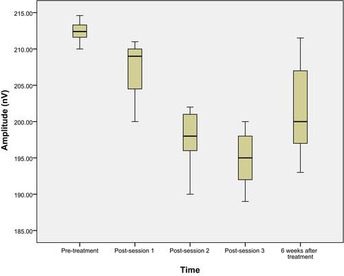
Table 3 The Overall Amplitude Changes in Percentage After Each Laser Session and 1.5 Months After Final Treatment Compared to Pretreatment Values
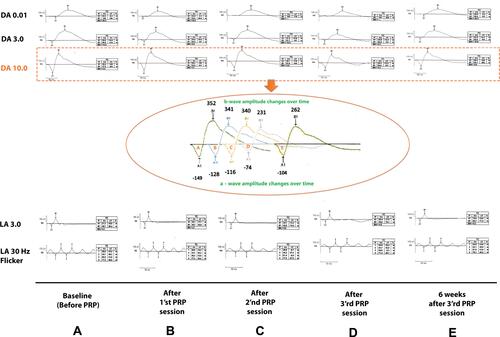
Figure 2 Full-field ERG of a patient enrolled in this study according to ISCEV −2015 standards. The time of ERG recording from baseline (before PRP) to 6 weeks after the final session of PRP has been shown in columns (A–E). Three upper rows are related to dark-adapted states (DA) and 2 lower rows are related to light-adapted states (LA) and between them magnified view of DA 10.0 wave changes of this patient over time have been shown in an ellipsoid inset. As have been shown, a- and b-waves amplitude decrease from baseline to 3ʹrd session after PRP (A–D) and these amplitudes recover and increase 6 weeks after the final session of PRP (E).