140
Views
3
CrossRef citations to date
0
Altmetric
Original Research

Vitreous and Serum Concentrations of Vascular Endothelial Growth Factor and Platelet-Derived Growth Factor in Proliferative Diabetic Retinopathy

, ORCID Icon, & ORCID Icon
Pages 1547-1552 | Published online: 09 Jun 2020

Figures & data

Table 1 Patients’ Characteristics

Table 2 The VEGF-A and PDGF-AB Concentrations

Table 3 The VEGF-A and PDGF-AB Concentration Ratios on Vitreous and Serum

Figure 1 The Vitreous VEGF-A and PDGF-AB concentrations in each PDR group.

Notes: Group classifications, I: PDR with VH; II: PDR with VH and fibrotic tissues; III: PDR with TRD. *Significant difference using Post hoc Bonferroni test (P<0.05).

Abbreviations: PDR, proliferative diabetic retinopathy; VH, vitreous hemorrhage; TRD, tractional retinal detachment; VEGF-A, vascular endothelial growth factor-A; PDGF-AB, platelet-derived growth factor-AB.

Figure 1 The Vitreous VEGF-A and PDGF-AB concentrations in each PDR group.Notes: Group classifications, I: PDR with VH; II: PDR with VH and fibrotic tissues; III: PDR with TRD. *Significant difference using Post hoc Bonferroni test (P<0.05).Abbreviations: PDR, proliferative diabetic retinopathy; VH, vitreous hemorrhage; TRD, tractional retinal detachment; VEGF-A, vascular endothelial growth factor-A; PDGF-AB, platelet-derived growth factor-AB.

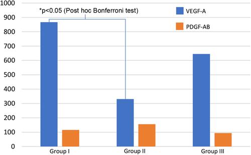
Table 4 The VEGF-A and PDGF-AB Concentration Ratios in Vitreous and Serum in Each PDR Group