
Figures & data
Figure 1 Stereoacuity of 40 patients after successful closure of a macular hole (MH) by MH surgery with internal limiting membrane (ILM) peeling. (A) Titmus stereoacuity of 40 patients with unilateral MH after surgery. Thirty-five eyes (87.5%) were considered to have normal stereoacuity of 40”-100” and 25 (62.5%) to have better stereoacuity of 40”-60”. (B) TNO stereoacuity for 40 patients with unilateral MH after surgery. Thirty-six eyes (90%) were considered normal (60”- 240”) and 22 (55%) were better (60”-120”).
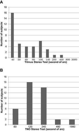
Figure 2 Correlation between age and postoperative stereopsis. (A) Titmus stereoacuity and (B) TNO stereoacuity.
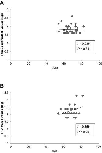
Figure 3 Correlation between size of macular hole (MH) and postoperative stereopsis. (A) Titmus stereoacuity and (B) TNO stereoacuity.
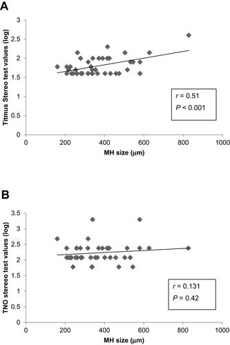
Figure 4 Correlation between postoperative best-corrected visual acuity and stereoacuity at more than 6 months after surgery. (A) Titmus stereoacuity and (B) TNO stereoacuity.
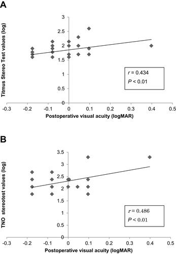
Table 1 Correlations Between Stereoacuity and Retinal Thickness and Foveal Shift Toward the Disc