Figures & data
Table 1 Demographic and Clinical Characteristics of Participants in Both Groups of the Study
Table 2 Comparison of the Recurrence Rate (PTT) Between Both Study Groups at Different Points of Follow-Up
Figure 1 Preoperative photograph of a patient with lower eyelid TT (A). At 1 week postoperatively with the lid margin in a good position without trichiasis (B). At 6 months with recurrent trichiasis (C).
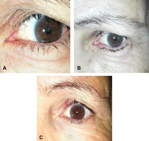
Figure 2 (A) Postoperative photograph of a patient with recurrent trichiasis and anterior lamellar laxity following ALR. (B) Postoperative photograph of another patient with recurrent trichiasis despite the lid margin in a good position following the same procedure.
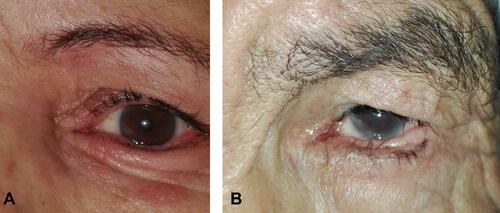
Figure 3 Cumulative survival analysis revealed that the mean time for recurrence is 8.8 months in the PTLR group (blue curve), while the mean time for recurrence for the ALR group (red curve) is 11.4 months.
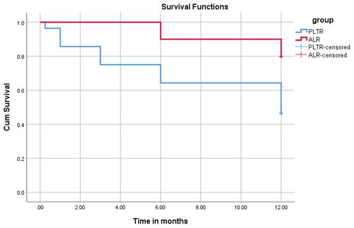
Table 3 Cosmetic Satisfaction of Participants in Each Group
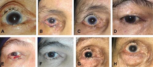
Figure 4 Pre- and postoperative photographs. (A) Preoperative photograph of a patient with a marginal entropion of the central one third of the lower eyelid, and postoperative appearance of the patient at 1 week (B) and 3 months (C) after anterior lamellar recession. (D) Preoperative photograph of another patient with lower eyelid marginal entropion with trichiasis and postoperative appearance at 1 week (E) and 12 months (F) following the same surgical procedure. (G) Preoperative appearance of an elderly woman with lower lid trichiasis without entropion compared to her postoperative appearance at 9 months (H).