Figures & data
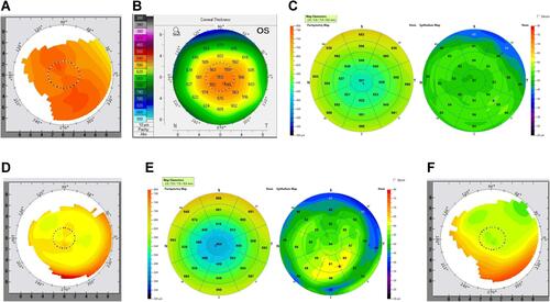
Figure 1 (A and B) Case 1. Pre-op topography and Pentacam pachymetry map. Note the lateralized central thin point relative to the apex, and pre-operative astigmatism. (C) Case 1. Pre-op OCT pachymetry and ETM. Overall relatively regular central epithelial thickness. (D) Case 1. 5 months post-op topography. Note the elliptical, irregular ablation pattern. (E) Case 1. 5 months post-op OCT pachymetry and ETM. Note the irregular thickening of the epithelium at the periphery of the ellipse. (F) Case 1. 2 months post-enhancement topography.
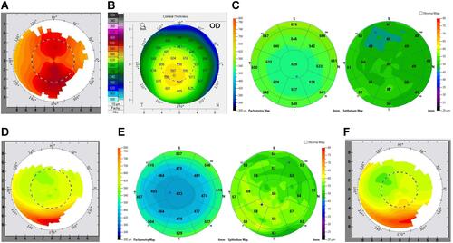
Figure 2 (A and B) Case 2. Pre-op topography. Note the lateralized thinnest point of cornea relative to the apex, and the pre-operative astigmatism. (C) Case 2. Pre-Op OCT pachymetry and ETM. Note the regular, mild variation of epithelial thickness. (D) Case 2. 5 months post-op. Note the elliptical irregular ablation. (E) Case 2. 5 Month Post Pachymetry and ETM. Note the irregular thickening of the epithelium at the periphery of the ellipse. (F) Case 2. 3 months post-enhancement.
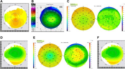
Figure 3 (A and B) Case 3. Pre-op Topography and Pentacam pachymetry. Note the lateralized thinnest point of cornea relative to the apex, and the pre-operative astigmatism. (C) Case 3. Pre-Op OCT Pachymetry and ETM. (D) Case 3. 5 month post-op. Note the elliptical, irregular ablation pattern. (E) Case 3. 5 Month Pachymetry and ETM. Note the irregular thickening of the epithelium of the elliptical. (F) Case 3. 3 months post enhancement. Enhancement done via topographic-guided ablation. Notice the smooth, round, more regular ablation pattern.
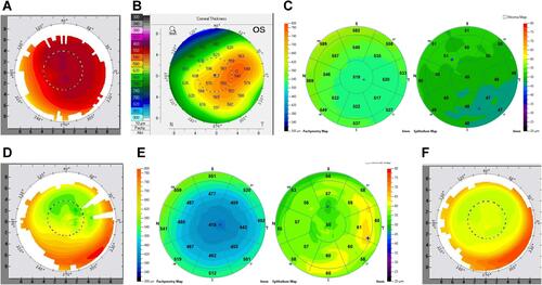
Figure 4 (A and B) Case 4. Pre-op topography and Pentacam pachymetry. Note the lateralized thinnest point of cornea relative to the apex, and the pre-operative astigmatism. (C) Case 4. Pre-Op OCT Pachymetry and ETM. Note the relatively regular central epithelial thickness. (D) Case 4. 5 months post-op. Note the elliptical, irregular ablation pattern. (E) Case 4. 5 Month Post Pachymetry and OCT. Note the irregular epithelial thickening at the periphery of the ellipse. (F) Case 4. 8 months post enhancement.
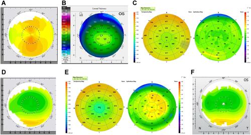
Figure 5 (A and B) Case 5. Pre-op topography and Pentacam pachymetry. Note the lateralized thinnest point of cornea relative to the apex. This is lateralized less than some of the other cases. The astigmatism here is also more irregular appearing, and the irregularity does correlate with the lateralized thin point. (C) Case 5. Pre-op OCT pachymetry and ETM. Note the relatively regular central epithelial thickness. (D) Case 5. 3 months post-op. Note the elliptical, irregular ablation pattern. (E) Case 5. 3 Month Post Pachymetry and OCT. Note the irregular epithelial thickening at the periphery of the ellipse. (F) Case 5. 4 months post enhancement.
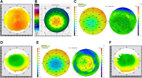
Figure 6 (A and B) Case 6. Pre-op topography and Pentacam pachymetry. Note the lateralized thinnest point of cornea relative to the apex, and the pre-operative astigmatism. (C) Case 6. Pre-op OCT pachymetry and ETM. Note the relatively regular central epithelial thickness. (D) Case 6. 2 months post-op. Note the elliptical shape and irregular ablation. (E) Case 6. 2 Month Post Pachymetry and OCT. Note the irregular epithelial thickening at the periphery of the ellipse. (F) Case 6. 10 months post enhancement. Note here that the elliptical shape is more exaggerated than 2 months post-op primary procedure likely due to continued epithelial thickening at the elliptical periphery.