
Figures & data
Table 1 Patient Demographics
Figure 1 Comparison of best-corrected visual acuity from baseline to final visit for each cohort and all studied eyes. *Statistically significant (p < 0.05).
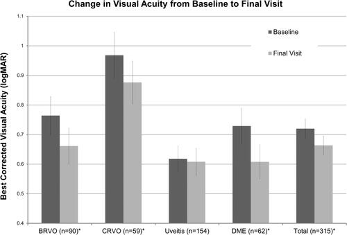
Figure 2 Comparison of central macular thickness on optical coherence tomography (OCT) from baseline to final visit for each cohort and all studied eyes. * Statistically significant (p < 0.05).
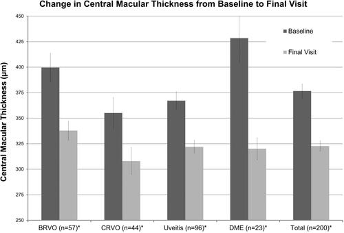
Figure 3 Change in BCVA compared to baseline following each DEX injection. Each graphed line includes only those patients who received at least the amount of DEX injections noted in the legend. Decreasing values signify improvement in BCVA. The BCVA values were recorded preferentially at the follow-up visit closest to six weeks after DEX injection and at least four weeks after treatment date. (A) All studied eyes, (B) BRVO, (C) CRVO, (D) DME, and (E) uveitis.
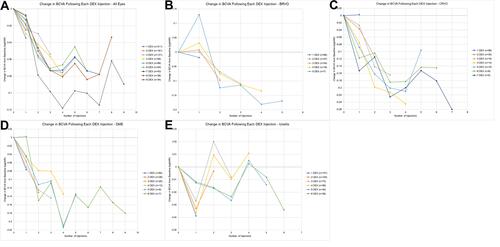
Figure 4 Comparison of baseline and final visit BCVA and CMT results for DME cohort based on lens status (pseudophakic throughout, phakic throughout, or underwent cataract surgery during study). Those eyes that underwent cataract surgery did so after an average of 1.6±0.2 DEX implants. The average change in BCVA and CMT was compared across each subgroup utilizing a Student’s t-test. Based on BCVA the following p-values were calculated: pseudophakic vs phakic –0.60, pseudophakic vs cataract surgery –0.83, and phakic vs cataract surgery –0.62. For CMT data the following p-values were calculated: pseudophakic vs phakic –0.09, pseudophakic vs cataract surgery –0.04, phakic vs cataract surgery 0.62.
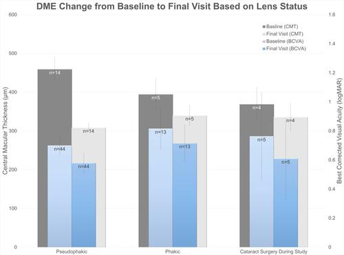
Table 2 Uveitis Cohort Supplemental Information
Figure 5 Uveitis cohort subgroup analysis with evaluation of the change in BCVA for various patient characteristics. Improvement in BCVA noted for the vitrectomized eyes, eyes without glaucoma diagnosis, age less than 60 years old, CMT greater than or equal to 400 µm and baseline visual acuity greater than or equal to 20/60. * Statistically significant (p < 0.05).
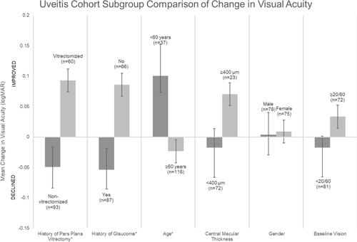
Figure 6 Best-corrected visual acuity in eyes undergoing cataract surgery compared from baseline (prior to initiation of intravitreal dexamethasone implant treatment) to the final visit. *Statistically significant (p < 0.05).
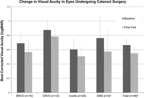
Table 3 Complications
Table 4 Maximum Intraocular Pressure