Figures & data
Figure 1 (A and B) Fundus photograph obtained using Cirrus™ HD-OCT before and after euthanasia, respectively. Optical coherence tomography (OCT) scanning includes the optic disk. (C) An OCT image obtained from a live rat. (D) The rat is placed on a custom-made platform, and the head and body are fixed.
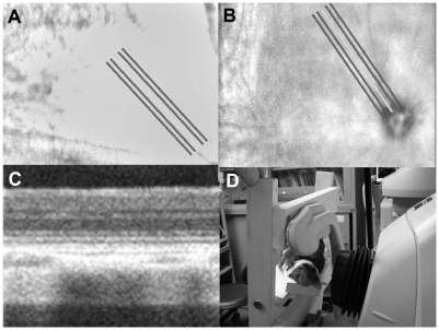
Figure 2 (A and B) The histopathology of the retina of two of four eyes after euthanasia. Even though artefacts due to the process of preparing specimens are observed, there are no obvious abnormalities in the retinal ganglion cell layer, inner plexiform layer, inner nuclear layer, outer plexiform layer, outer nuclear layer, or the photoreceptor inner and outer segments. (C and D) The magnified histology of the retina of two of four eyes after euthanasia corresponding to (A) and (B), respectively (magnification unknown). Photoreceptor inner (IS) and outer segments (OS) appear normal. The outer nuclear layer (ONL) looks normal.
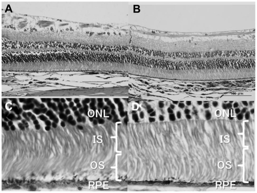
Figure 3 Optical coherence tomography (OCT) images of retina before (A–D) and after euthanasia (E–H corresponding to A–D, respectively). Figure 3A corresponds to . Figure 3D corresponds to . The internal limiting membrane line (white arrow), the outer plexiform layer (OPL) line (blank arrow head), the external limiting membrane (ELM) line (white arrow head), the inner and outer segments (IS/OS) (black arrow head), and the retinal pigment epithelium line (black arrow) on OCT images are observed in all four eyes prior to euthanasia (A–D). After euthanasia, the ELM (white arrow head) and the IS/OS (black arrow head) disappears in three of the four eyes (F–H) and, in one eye, the intensity of the ELM (white arrow head) and the IS/OS (black arrow head) is reduced (E). The OPL line (blank arrow head) is still observed in all four eyes (E–H).
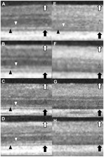
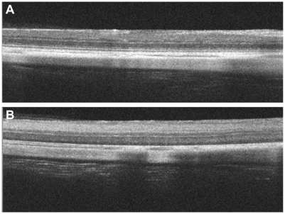
Figure 4 Optical coherence tomography images of the retina before (A) and after (B) euthanasia of 6-mm scan. Tomographic images of 6-mm-long show that neither the inner and outer segments nor the external limiting membrane are evident in a widespread area of rat retinas after euthanasia.