Figures & data
Figure 1 DFG epidermis removal techniques. (A) The donor site is marked (upper outer buttock quadrant). (B) #15 Bard-Parker blade dissection. (C) After complete removal of epidermis (multiple spot hemorrhages and scattered foci of exposed fat). (D) The dermis fat graft following epidermal scalpel dissection. (E) Debridement of the graft epidermis using the bipolar diathermy. (F) The dermis fat graft after electrocoagulation of the epidermis.
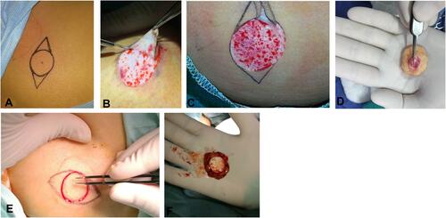
Table 1 Baseline Characteristics of Patients in Both Groups
Figure 2 (A) Preoperative photo of a patient with congenital microphthalmia. (B) Postoperative appearance with complete healing and mild discharge 8 week after surgery (Scalpel dissection). (C) After prosthesis fitting with adequate cosmesis despite of lower lid mild entropion.
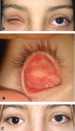
Figure 3 Complete epithelialization at postoperative week 8 with hair growth following epidermal scalpel dissection.
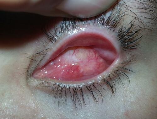
Figure 4 (A) Preoperative appearance of right phthisis bulbi with marked enophthalmos and poor prosthesis retention. (B) Complete healing at postoperative week 16 (Scalpel dissection). (C) After prosthesis fitting with good volume replacement. Lateral tarsal strip was performed.
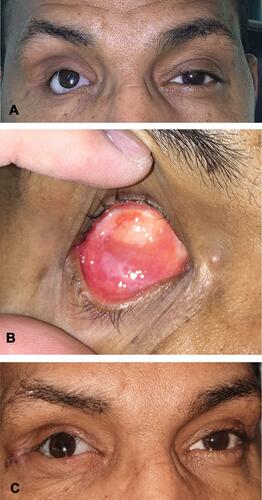
Table 2 Postoperative Outcome
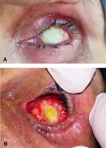
Figure 5 (A) Postoperative view of a patient in the electrocoagulation group at the end of the surgery. (B) Dermal ulceration noticed 8 weeks postoperatively. (C) Complete spontaneous healing after 32 weeks.
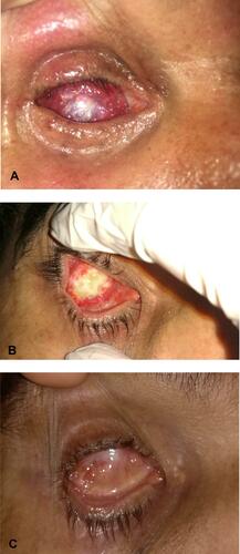
Figure 6 (A) Postoperative photograph shows central dermal ulceration 10 weeks postoperative (electrocoagulation). (B) After complete healing (after 28 weeks). (C) The prosthesis is fitted before complete healing.
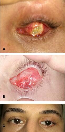
Figure 7 (A) Postoperative photograph shows persistent white color of the dermal surface after electrocoagulation at postoperative week 8 that ended up with persistent ulcer (B). Excision with amniotic membrane transplantation was performed for this patient.