Figures & data
Table 1 Demographics
Table 2 The Type and Magnitude of Residual Refractive Errors for Each of the 5 Categories
Figure 1 (A) Pre-op topography. (B) Pre-op pachymetry showing corneal thickness. (C) Pre-op OCT pachymetry. (D) Pre-op OCT epithelial thickness mapping (ETM). (E) 5 month post-op topography. (F) 5 month post-op OCT pachymetry. (G) 5 month post-op OCT ETM- note the irregular epithelial thickening at the edge of the epithelial ablation.
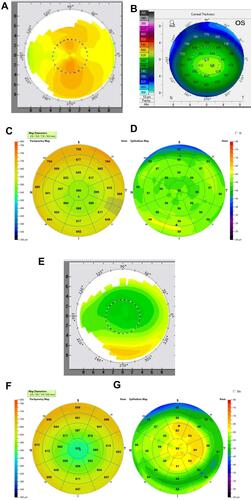
Figure 2 (A) Pre-op pachymetry showing corneal thickness.(B) Pre-op epithelium map. – 9 mm ETM (Avanti XR).
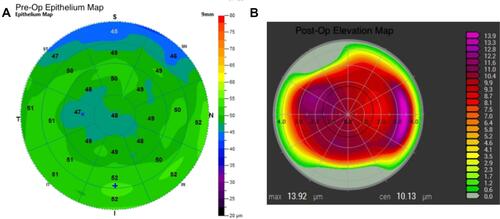
Figure 3 (A) Pre-op topography (Topolyzer Vario) (B and C) Pre-op OCT pachymetry and epithelial thickness map. 9mm ETM (Avanti XR), (D) 1 month post-op topography, (E and F) 1 month post-op OCT pachymetry and epithelial thickness map. 9mm ETM (G) Post-enhancement 1 year.
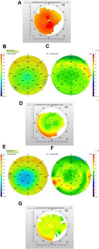
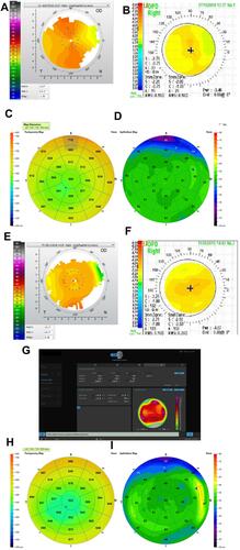
Figure 4A Continued.
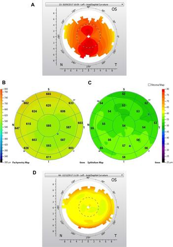
Figure 4 (A) Pre-op topography. (B) Pre-op OCT pachymetry. (C) Pre-op epithelial thickness map. (D) Post-op 1 month topography. (E) Post-op 1 month OCT pachymetry. (F) 1- month post-op epithelial thickness map 9mm ETM (Avanti XR). The thickened epithelium as opposed to pre-op. (G) Post-enhancement and 1 year post-op. (H, I) OCT Pachymetry map post enhancement and 1 year post-op.
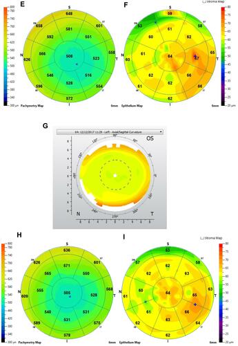
Figure 5 (A) Pre-op topography map (Topolyzer Vario). (B) Pre-op pachymetry showing corneal thickness. (C) Pre-op epithelial thickness map. 9mm ETM (Avanti XR). (D) 3-month post-op topography. (E) 3-month post-op OCT pachymetry (F) 3-month post-op epithelial thickness map (ETM). (G) 4-month post ENH topography. (H) 4-month Post ENH OCT pachymetry. (I) 4-month Post ENH epithelial thickness map (ETM).