Figures & data
Figure 1 Pintucci KPro.
Notes: (A) Pintucci KPro device. (B) First stage – insertion of Pintucci device into submuscular pouch for vascular ingrowth. (C) First stage – covering the ocular surface with buccal/labial mucosa. (D) Second stage – implantation of the retrieved device into the eye (2–3 months after the stage 1). (E) Second stage – creating an opening in the buccal mucosa to expose the optic. (F) Postoperative appearance of a successful Pintucci KPro. All pictures of this figure are the courtesy of Dr Qureshi Maskati.
Abbreviation: KPro, keratoprosthesis.
Abbreviation: KPro, keratoprosthesis.
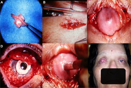
Table 1 Historical kertaoprostheses
Figure 2 AlphaCor keratoprosthesis.
Notes: (A) AlphaCor device. (B) First stage – insertion of AlphaCor within the corneal lamellar pocket. A 3 mm central zone part of the posterior lamella is trephined. (C) Second stage – the external portion of the optic is exposed by excision of the superior corneal lamella. Adapted by permission from Macmillan Publishers Ltd: Eye. Hicks CR, Crawford GJ, Lou X, et al. Corneal replacement using a synthetic hydrogel cornea, AlphaCor[trade]: device, preliminary outcomes and complications. 2003;17(3):385–392, Copyright ©2003.Citation109
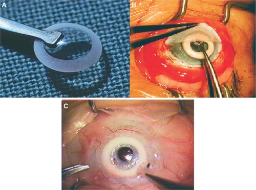
Figure 3 Boston keratoprosthesis.
Notes: (A) Type-1 device with front plate (optical stem) and titanium back plate. (B) Type-1 device in situ with a bandage contact lens. (C) Type-2 device with extended optical stem and titanium back plate. (D) Type-2 device in situ – projection of the optical stem through the lid. All pictures of this figure are the courtesy of Dr James Chodosh.
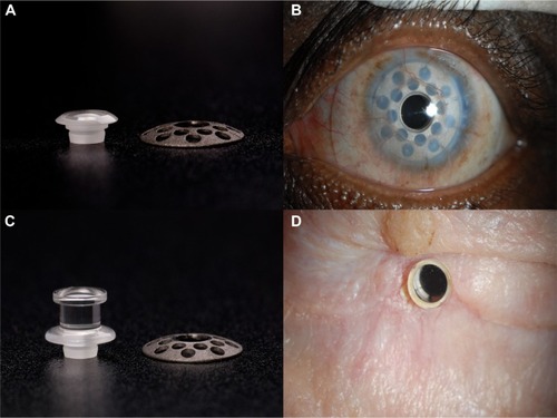
Figure 4 Assembly and implantation of Boston type-1 KPro.
Notes: (A) Donor corneal button is trephined to create a central aperture. (B) Corneal graft is placed on the front plate. (C) PMMA back plate covers the graft and titanium ring locks the device. (D) Assembled type-1 device ready for implantation (corneal graft is sandwiched between the front and back plates). (E) Host cornea is excised (excised diameter matches outer diameter of the corneal graft). (F) Implantation of the type-1 KPro. All pictures of this figure are the courtesy of Dr Geetha Iyer.
Abbreviations: KPro, keratoprosthesis; PMMA, polymethylmethacrylate.
Abbreviations: KPro, keratoprosthesis; PMMA, polymethylmethacrylate.
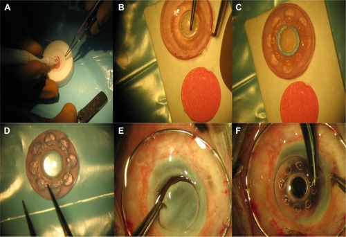
Figure 5 Postoperative course of the Boston type-1 KPro.
Notes: (A) Corneal graft melt adjacent to the front plate. (B) Retroprosthetic membrane seen through the optic. (C) OCT scan showing Boston type-1 KPro in situ. Pictures (A) and (B) are courtesy of Dr Geetha Iyer.
Abbreviations: KPro, keratoprosthesis; OCT, optical coherence tomography.
Abbreviations: KPro, keratoprosthesis; OCT, optical coherence tomography.
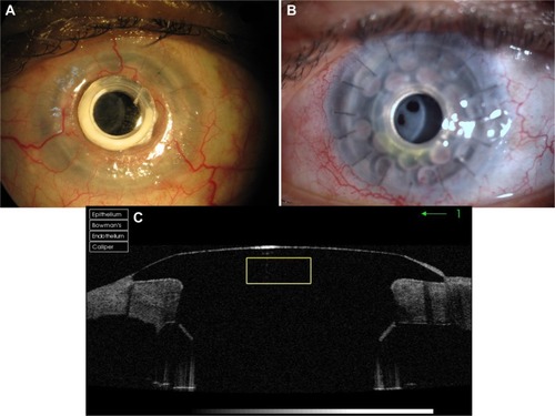
Figure 6 OOKP stage 1 – buccal mucosal graft preparation and transplantation.
Notes: (A) Excision of the BMM. (B) Excised mucosal tissue-fat and muscle are trimmed off. (C) Ocular surface preparation (sclera is bared and corneal epithelium debrided). (D) Mucosal graft transplantation on to the ocular surface.
Abbreviations: OOKP, osteo-odonto-keratoprosthesis; BMM, buccal mucous membrane.
Abbreviations: OOKP, osteo-odonto-keratoprosthesis; BMM, buccal mucous membrane.
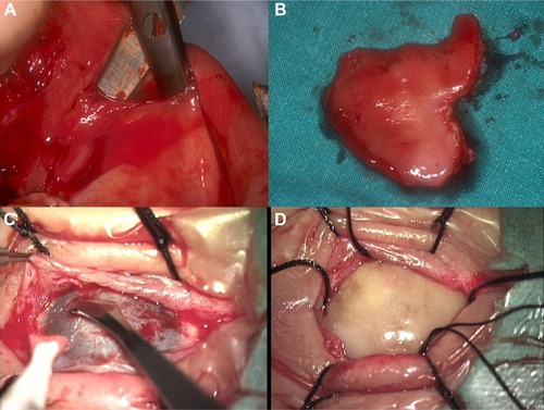
Figure 7 OOKP stage 1 – tooth extraction and preparation of the OOAL lamina.
Notes: (A) Extraction of the tooth with a piece of mandible. (B) Preparation of the tooth (dentine is exposed on one surface, while the alveo-dental ligament is preserved). (C) A central hole is drilled perpendicular to the lamina. (D) A PMMA cylinder is inserted into the lamina.
Abbreviations: OOKP, osteo-odonto-keratoprosthesis; OOAL, alveo-dento-acrylic; PMMA, polymethylmethacrylate.
Abbreviations: OOKP, osteo-odonto-keratoprosthesis; OOAL, alveo-dento-acrylic; PMMA, polymethylmethacrylate.
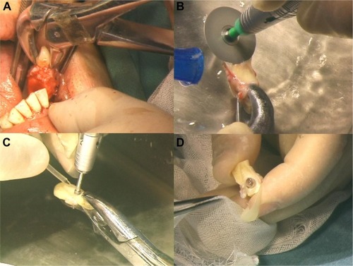
Figure 8 OOKP lamina and postoperative course after stage 1.
Notes: (A) PMMA optical cylinder – the wide part sits on the dentine surface. (B) Prepared OOAL lamina – rim of acrylic cement can be seen around the optic. (C) A healthy mucous membrane 1 month after the stage 1.
Abbreviations: OOKP, osteo-odonto-keratoprosthesis; PMMA, polymethylmethacrylate; OOAL, alveo-dento-acrylic.
Abbreviations: OOKP, osteo-odonto-keratoprosthesis; PMMA, polymethylmethacrylate; OOAL, alveo-dento-acrylic.
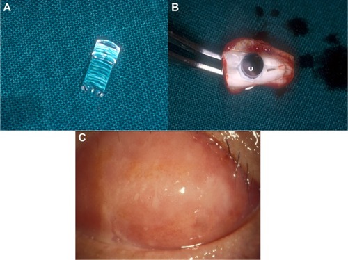
Figure 9 OOKP stage 2 – retrieval of the lamina and implantation into the eye.
Notes: (A) Lamina is recovered from the subcutaneous pocket. (B) Connective tissue is removed from the lamina to expose the dentine side. (C) Buccal mucosa from the eye is reflected to expose the cornea. (D) Central corneal button is excised. (E) Lens extraction (IOL is also removed when present). (F) Open sky core vitrectomy is performed. (G) Lamina is implanted into the eye with wide portion of the optical stem passing through the cornea. (H) Mucosal membrane is replaced over the lamina. Through a central opening in the BMM, the optic projects beyond 1 mm.
Abbreviations: OOKP, osteo-odonto-keratoprosthesis; BMM, buccal mucous membrane; IOL, intraocular lens.
Abbreviations: OOKP, osteo-odonto-keratoprosthesis; BMM, buccal mucous membrane; IOL, intraocular lens.
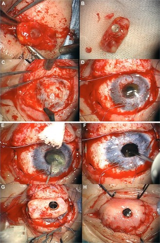
Figure 10 Postoperative course after stage 2.
Notes: (A) A healthy OOKP eye after a successful surgery. (B) CT scan image showing an intact lamina in the OOKP eye.
Abbreviations: OOKP, osteo-odonto-keratoprosthesis; CT, computerized tomography.
Abbreviations: OOKP, osteo-odonto-keratoprosthesis; CT, computerized tomography.
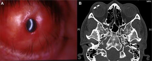
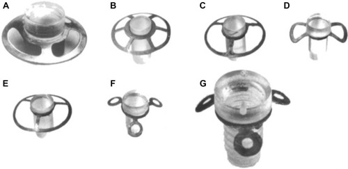
Figure 11 Filatov keratoprostheses.
Notes: (A–F) show the previous models. Model (G) is the latest and most successful Iakymenko prosthesis. Reproduced from Iakymenko S. Forty-five years of keratoprosthesis study and application at the Filatov Institute: a retrospective analysis of 1060 cases. Int J Ophthalmol. 2013;6(3):375–380, doi:10.3980/j.issn.22223959.2013.03.22.Citation106