Figures & data
Figure 1 Appearance of asteroid hyalosis in slit-lamp with direct focal illumination (A) and retinal examination here seen using a fundus photography (B).
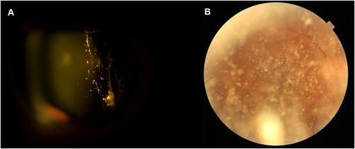
Figure 2 Asteroid hyalosis is seen as numerous hyperreflective foci in the vitreous in cross-sectional optical coherence tomography (OCT) scans (A) as well as 3D OCT (B), and as small round hyperechoic elements in the vitreous in B-scan ultrasonography (C).
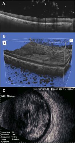
Table 1 Differential Diagnoses of Asteroid Hyalosis
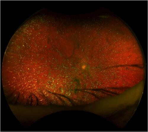
Figure 3 Scanning laser ophthalmoscopy-based systems such as OPTOS may enable retinal examination to a certain degree.