Figures & data
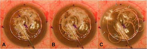
Figure 1 Type 1 big bubble formation with stromal whitening. (A) Stromal whitening extending from the site of air injection to the limbus on one side. (B) Central commencement of Type 1 bubble (black arrows) with displacement of the anterior chamber (AC) bubbles to the periphery (white arrows). (C) Complete formation of Type 1 bubble.
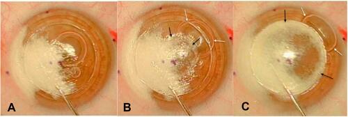
Figure 2 Incomplete Type 1 big bubble formation without stromal whitening. (A) Central commencement of Type 1 bubble in a clear cornea (black arrows). (B and C) Enlargement of Type 1 bubble (black arrows) with AC bubble displacement to the periphery (white arrows).
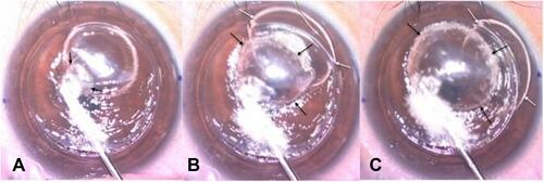
Figure 3 Type 2 bubble formation after a small intrastromal bubble. (A) Air injection with the formation of a small intrastromal bubble (black arrows). (B) Rapid formation of Type 2 bubble as a small central bubble (white arrows). (C) Centrifugal enlargement of Type 2 bubble (white arrows).
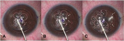
Figure 4 Midperipheral formation of Type 2 bubble after a small intrastromal bubble. (A) Air injection with the formation of a small intrastromal bubble (black arrows). (B and C) Formation of Type 2 bubble enlarging inferiorly (white arrows).
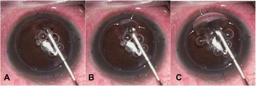
Figure 5 Direct access of air to the plane between the DM and stroma to form Type 2 bubble. (A and B) Central commencement of Type 2 bubble (white arrows). (C) Centrifugal enlargement of Type 2 bubble (white arrows).
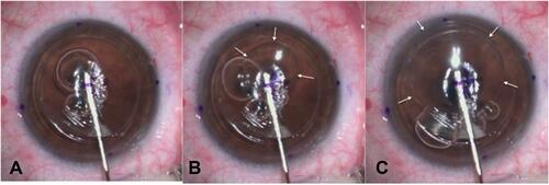
Figure 6 Mixed bubble without stromal whitening. (A) Central commencement of Type 1 bubble (black arrows). (B) Enlargement of the bubble towards one side (black arrows). (C) Rapid formation of type 2 bubble (white arrows).