Figures & data
Figure 1 Design of extended IBEB. The medial end of the excised skin pad is 5 mm laterally in from the start of the eyebrow, and the lateral end of the skin pad is 5–10 mm laterally out from the tail of the eyebrow. The incision is made perpendicularly to the hair shafts. The vertical width of the skin pad is 10–20 mm at its widest point in most patients.
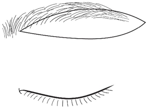
Figure 2 Planning the desirable postoperative eyebrow shape. The desirable eyebrow shape after surgery is shown (gray area). Postoperative descent of the eyebrow, which is generally observed after extended IBEB, has to be considered. It is unnecessary to transplant hair into the infrabrow operative scar (dotted line), as it would be covered by the regrowth of hair, made possible by incision perpendicular to the hair shafts.
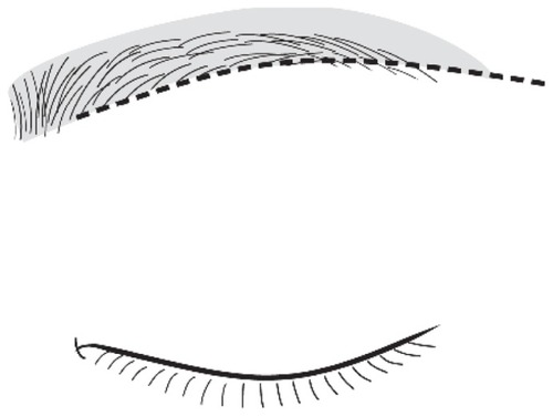
Figure 3 Marking of the area of hair transplantation. The area above the upper margin and the tail of the eyebrow (shadow area) is designed so that it will develop into the desired shape.
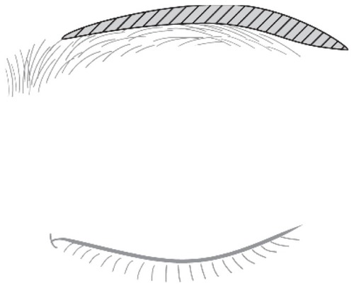
Figure 4 Case 1. (A) A 50-year-old man with moderate bilateral dermatochalasis. (B) Extended IBEB with FUT was carried out. The maximum width of the skin pad was 17 mm. The subseptal fat was dissected. The incision was made perpendicularly to the hair shafts. The numbers of follicular units available to transplant were 100 on each side, and these were transplanted in the upper part of the eyebrow from body to tail. (B′) The portion of excised the eyebrow, is area A. Approximately half of the eyebrow was excised from the body to the tail of the eyebrow. The portion of removed orbital fat is area B. The portion of transplanted hair to the eyebrow is area C. It extended from the center of the eyebrow to the outside of the eyebrow’s superior border with a maximum width of 7 mm. (C) Twelve months after the operation, the scar had become less conspicuous. The scar just along the lower margin of the eyebrow was partially covered by regrown hair. The transplanted hair was growing. (D) Twenty-four months after the operation, the regrown eyebrow entirely covered the scar and made it inconspicuous. The transplanted hair was also fully grown. Any changes in, reduction, or distortion of the eyebrow were acceptable. The patient was satisfied with the result and no complications were observed. He opened his eyes more easily, his visual field had become larger, and his lid creases had a more youthful appearance.
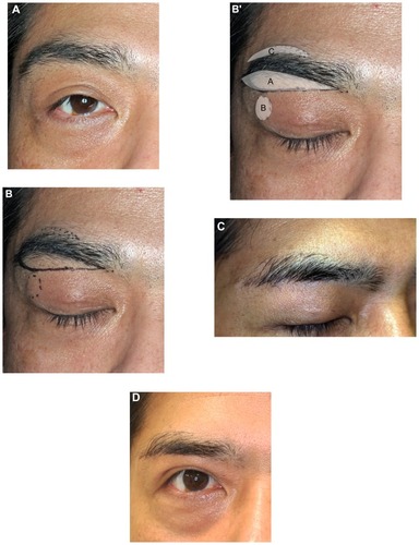
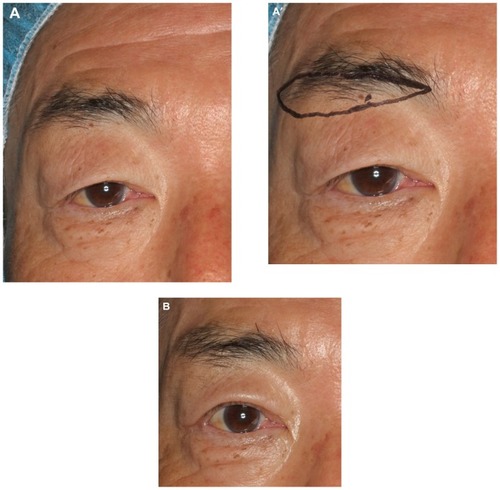
Figure 5 Case 2. (A) A 56-year-old man with severe bilateral dermatochalasis. He had a sleepy looking appearance due to droopy eyelids. Lateral hooding impaired the upper visual field. The maximum width of eyebrows was 1 cm. (A′) Extended IBEB with FUT was carried out, where the maximum width of the excised skin pad was 20 mm. The numbers of follicular units available to transplant were 150 on each side, and these were transplanted into the upper part of the eyebrow from body to tail. (B) Twelve months after the operation, the decline and reduction of the eyebrow was slight and the patient was able to open his eyes easily. The maximum width was not as variable as before the operation at 1 cm. He was satisfied with the result and no complications were observed.