Figures & data
Table 1 Comparison of Primary Cancer Sites in Different Cohorts
Table 2 Comparison of Primary Cancer Site Against Sex
Table 3 Segmentation of Primary Cancer Site by Age
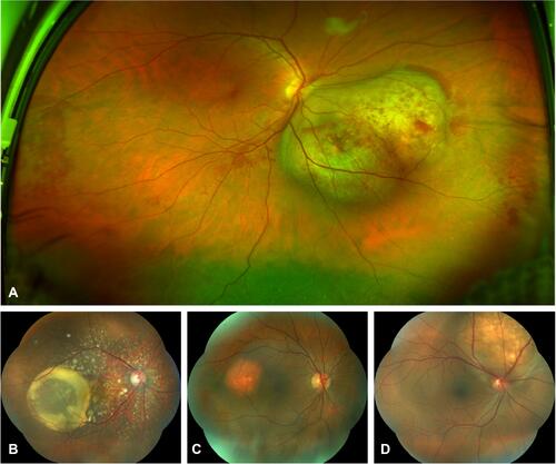
Figure 1 (A) Ultra-wide field fundus image of a single lesion, bi-lobulated, dome-shape choroidal metastasis from lung cancer, with intraretinal microhemorrhages over and around the tumor, as well as subretinal fluid in the lower retina quadrants. (B) Choroidal metastasis from breast cancer with multiple, disseminated seeds and a large cystoid subretinal fluid accumulation. (C) Renal cancer metastasis, reddish-orange, slightly elevated and round tumor. (D) Solitary, white-yellowish lesion from pulmonary cancer with subretinal fluid around the metastasis.