
Figures & data
Figure 1 Treatment protocol. Intervention schedule for patients with pachychoroid neovasculopathy and neovascular age-related macular degeneration. 
 clinical data collection;
  efficacy endpoint.
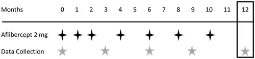
Table 1 Patient Characteristics at Baseline and 12 Months After Intravitreous Injection of Aflibercept
Table 2 Comparison of Baseline Characteristics Between Patients with Pachychoroid Neovasculopathy and Neovascular AMD
Figure 2 A case with pachychoroid neovasculopathy (before treatment). Images of the left eye of a 70-year-old man with pachychoroid neovasculopathy without any treatment history. The best-corrected visual acuity was 20/50. (A) Color fundus photograph shows subretinal hemorrhage and serous retinal detachment. Note that drusen are absent. (B) Optical coherence tomography (OCT) angiography image shows choroidal neovascularization (CNV). (C) Late-phase fluorescein angiography image shows leakage suggesting occult CNV. (D) Late-phase indocyanine green angiography image. (E) Enhanced-depth imaging OCT image shows serous retinal detachment and elevation of the retinal pigment epithelium, suggesting CNV. The choroid is thick throughout the macula, and choroidal vessels are dilated. Subfoveal choroidal thickness = 387 μm, central retinal thickness = 317 μm.
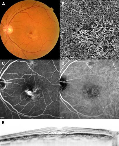
Figure 3 A case with pachychoroid neovasculopathy (after treatment). Images of the same patient as in , acquired 12 months after initial treatment. The best-corrected visual acuity had improved to 20/16. (A) Color fundus photograph shows resolution of subretinal hemorrhage and serous retinal detachment. (B) Optical coherence tomography (OCT) angiography image shows choroidal neovascularization. (C) Late-phase fluorescein angiography image shows reduction of leakage. (D) Late-phase indocyanine green angiography image. (E) Enhanced-depth imaging OCT image shows resolution of serous retinal detachment. Subfoveal choroidal thickness = 348 μm, central retinal thickness = 206 μm.
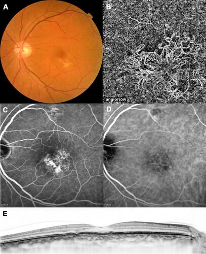
Table 3 Changes in Visual Acuity, Subfoveal Choroidal Thickness, and Central Retinal Thickness During Intravitreal Aflibercept Injection Treatment for Pachychoroid Neovasculopathy
Table 4 Changes in Visual Acuity, Subfoveal Choroidal Thickness, and Central Retinal Thickness During Intravitreal Aflibercept Injection Treatment for Neovascular Age-Related Macular Degeneration
Table 5 Patients Classified with Pachychoroid Neovasculopathy
Table 6 Prevalence of Dry Macula in Patients with Pachychoroid Neovasculopathy and AMD Treated with Aflibercept