
Figures & data
Table 1 Demographic Characteristics of Patients with Ocular Malignancies (N = 246)
Table 2 Clinicopathological Characteristics of Patients (N = 246)
Table 3 Pathological Diagnoses According to Malignancy Type (N = 246)
Figure 1 (A) Clinical photograph of a 80-year-old female patient with a left upper eyelid nodulo-ulcerated sebaceous gland carcinoma with preauricular lymph node metastasis. (B) Microphotograph of a moderately differentiated sebaceous gland carcinoma showing lobules of malignant cells with sebaceous differentiation.
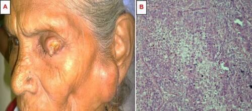
Figure 2 (A) Clinical photograph of a pigmented basal cell carcinoma on the left lower eyelid. (B) Microphotograph of a basal cell carcinoma showing nests of atypical pigment-laden basal cells with peripheral palisading and mitotic figures.
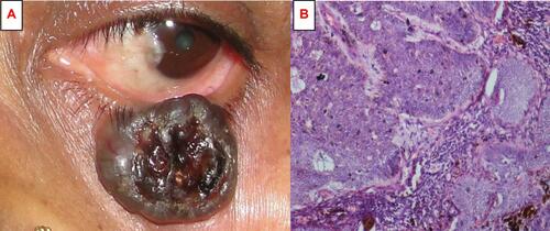
Figure 3 (A) Clinical photograph of a male child with an orbital retinoblastoma on the right eye. (B) Computed tomography in the sagittal view showing intracranial extension with lymph node and cerebral metastasis.
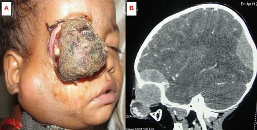
Figure 4 (A) Clinical photograph of a 15-year-old male patient with an orbital rhabdomyosarcoma in the left superior rectus muscle of the eye showing non-axial proptosis, marked chemosis and exposure keratopathy. (B) Computed tomography in the coronal view showing a large, round, well-defined, enhancing mass over the superior orbit, pushing the globe inferiorly. (C) Clinical photograph after five cycles of systemic neoadjuvant chemotherapy (vincristine, doxorubicin, and cyclophosphamide) showing complete regression of tumor and alopecia. (D) Immunohistochemical stains reveal positivity for vimentin within the cytoplasm of rhabdomyosarcoma cells.
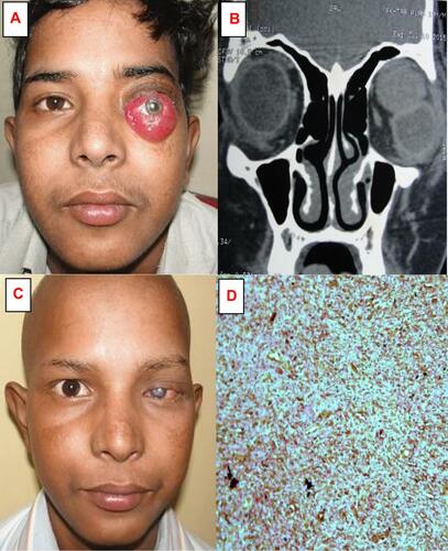
Figure 5 (A) Clinical photograph of a giant ocular surface squamous neoplasia (about 250°) in the right eye. (B) Clinical photograph after four cycles of topical 5FU drops.
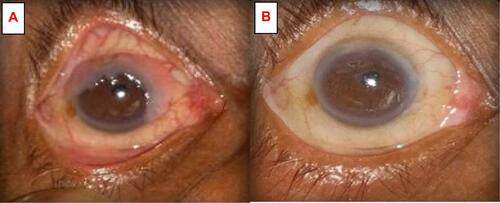
Table 4 Histopathological Differentiation of Eyelid Malignancies (N= 92)
Table 5 Distribution of Cases According to Treatment Procedures Undertaken by Patients (N= 246)
Table 6 Distribution of Study Subjects According to Treatment Outcomes After Six Months (N = 246)