
Figures & data
Figure 1 Drawing of the elastic silicone sheet.
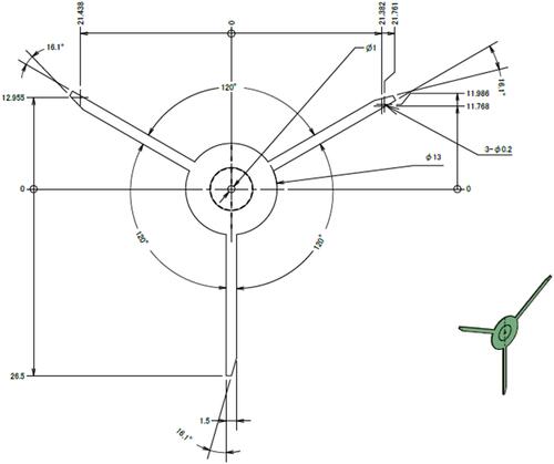
Figure 2 Surgical procedure of a case with a subluxated lens. (A) One arm was inserted into the corneal side port using forceps. (B) The elastic silicone sheet (ESS) was inserted into the eye behind the subluxated lens through a 3-mm corneoscleral incision and zonular dialysis. (C) The other two ends of the arms were inserted through the corneal side ports. (D) Phacoemulsification and aspiration (PEA) was performed on the ESS. (E) After the PEA was completed, the two arms were dissected. (F, G) The remaining arm was caught and pulled out. (H) The ESS was easily removed through the corneoscleral incision.
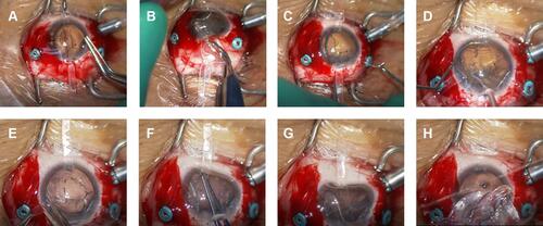
Table 1 A Summary of the Patients