
Figures & data
Figure 1 (A) Pre chemotherapy photograph of a 70 years old male patient having left lower eyelid ulceronodular type sebaceous gland carcinoma. (B) After 3 cycles of neoadjuvant chemotherapy (cis-platinum and 5 FU), showing partial response of chemotherapy.
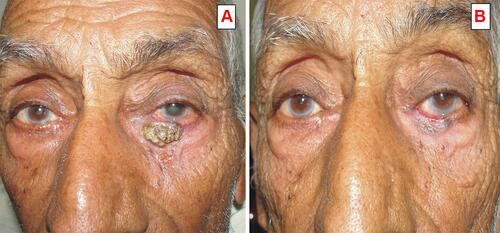
Figure 2 (A) Clinical photograph of a 48 years old male patient having right lower eyelid squamous cell carcinoma with orbital invasion before chemotherapy. (B) After 5 cycles of neoadjuvant chemotherapy and 3 cycles of external beam radiation.
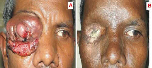
Figure 3 Microphotograph of a different histological section stained (Hematoxylin and Eosin x 100 (X) showing (A). Rhabdomyosarcoma round cells (primitive rhabdomyoblast). (B). Sebaceous gland carcinoma moderately differentiated showing lobules of malignant cells with sebaceous differentiation. (C). Basal cell carcinoma showing nests of pigment laden atypical basal cells with peripheral palisading and mitotic figures. (D). Squamous cell carcinoma showing keratin pearls, numerous malignant cells, and mitotic figure. (E). OSSN showing keratin pearls and proliferation of irregular dysplastic epithelium with infiltration of sub epithelial tissue (F). Retinoblastoma well differentiated with Flexner-Wintersteiner rosette arrangement.
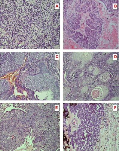
Table 1 Analysis for Serum Malondialdehyde (MDA) Levels in Controls (N=16) and Cancer Patients (N=32, Before and After Chemotherapy)
Table 2 Analysis for Serum Malondialdehyde (MDA) Levels According to Age of Patient and Tumor Advancement (Before and After Chemotherapy)
Table 3 Analysis for Serum Malondialdehyde (MDA) Levels According to Histopathological Type and Differentiation. (Before and After Chemotherapy)