
Figures & data
Table 1 Triggering Events in SO Patients
Figure 1 After the inciting event, the antigen-presenting cells (APC) present the antigen peptide to autoreactive CD4+ cells. This activates the CD4+ cells and brings about the autoimmune reaction damaging the other (sympathizing) eye.
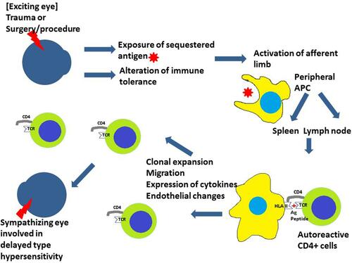
Figure 2 (A) Fundus photo of the posterior pole of the sympathizing eye shows a blurred margin of the optic disc, radial retinal folds around the optic disc, and subretinal fluid at the posterior pole. (B) FFA image at early venous phase shows disc leak, choroidal folds, and pinpoint leaks. (C) FFA image at mid-venous phase shows the typical multiple pinpoint leaks more clearly. (D) FFA image at late venous phase shows disc leak and blurred margins of pinpoint leaks.
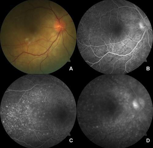
Figure 3 (A) Anterior segment photo shows a phthisical eye with evidence of repair of globe rupture. (B) Posterior segment photo of the other eye shows depigmented fundus (sunset glow) with areas of fibrotic pigmentary changes.
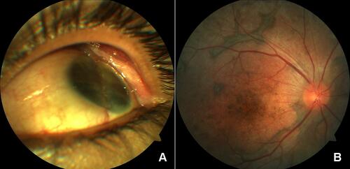
Table 2 Clinical Features of Patients with Sympathetic Ophthalmia (SO)
Table 3 The Spectrum of Various Immunomodulatory Agents Used in the Treatment of Sympathetic Ophthalmia (SO)