Figures & data
Table 1 Demographic, Preoperative BCVA, Postoperative BCVA and Functional Outcomes of the Different Groups
Table 2 Comparison Between Pre- and Postoperative BCVA of Different Groups of Macular Holes
Table 3 Anatomic and Functional Outcomes of Subgroups of Idiopathic Macular Hole
Table 4 Postoperative VA Stratified According to Macular Hole Status in Different Groups of Macular Holes
Figure 1 (A) High-definition 5-line raster OCT image of the right eye of a 66-year-old male with idiopathic FTMH. Note that the posterior hyaloid is fully detached along with an operculum (star). The hole edges are thickened with cystic spaces in a typical pregnant draw-bridge appearance. Both ends of the ELM and IS/OS junction layers are drawn upwards (2 white arrows). MLD of the hole was 216µ. Preoperative BCVA was 6/24. (B) Postoperative OCT image of the same patient taken at 1-month follow-up visit. Note U-type hole closure with restoration of ELM and IS/OS layers. There is a residual shallow sub-foveal neurosensory detachment (white arrow), Postoperative BCVA was 6/9.
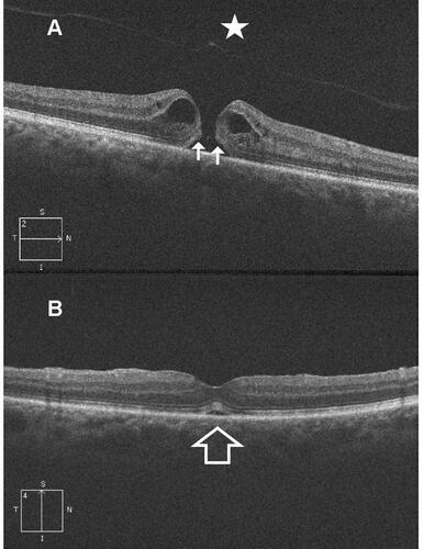
Figure 2 (A) High-definition 5-line raster OCT image of the left eye of a 75-year-old female with idiopathic FTMH. Note the fully detached posterior hyaloid and the hole operculum (star). The hole shows thickened edges with cystic spaces, and upward lift of the ELM and IS/OS junction layers (2 white arrows). MLD of the hole was 200µ. Preoperative BCVA was 6/36. (B) Postoperative OCT image of the same patient taken at 3-month follow-up visit. Note U-type hole closure with restoration of ELM and IS/OS layers. Note the residual defect in the IS/OS junction layer (white arrow head). Postoperative BCVA improved to 6/9.
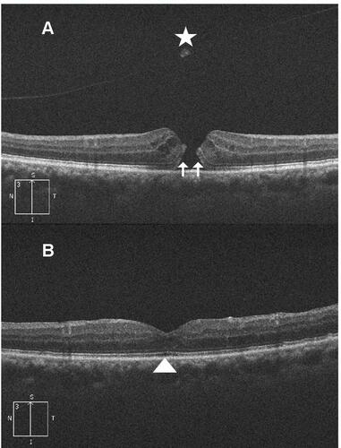
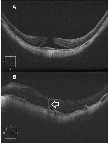
Figure 3 (A) High-definition 5-line raster OCT image of the left eye of a 46-year-old female with myopic FTMH. Note the OCT features of myopia as posterior bowing of the sclera, thinning of the choriocapillaris and dome-shaped macula. MLD of the hole was 328µ. Preoperative BCVA was 5/60. (B) Postoperative OCT image of the same patient taken at 1-month follow-up visit. The hole has closed though with interrupted ellipsoid zone. Note the hyporeflective intra-retinal cystic spaces. The hyperreflective vertical line traversing the neurosensory retina from the fovea to the RPE represents a track line which is a marker of previous injury to ELM layer and damage to the photoreceptors around the line (white arrow). The RPE is thickened and shows multiple pigment epithelial detachments. Postoperative BCVA remained 5/60.