Figures & data
Table 1 Contribution of Different Etiologies of Uveitis
Figure 1 Positive tuberculin reaction in a patient with tuberculous uveitis.
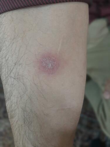
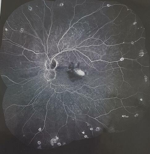
Figure 2 Intermediate uveitis and peripheral vasculitis in a patient with tuberculosis.
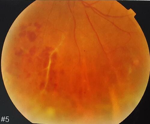
Figure 3 Vitiligo in a patient with Vogt–Koyanagi–Harada syndrome.
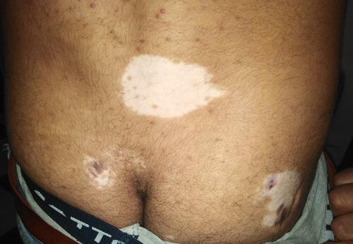
Figure 4 Multiple detachments in a patient with Vogt–Koyanagi–Harada syndrome.
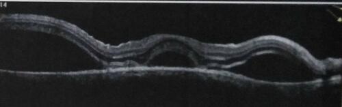
Figure 5 Active CNV in a patient with Vogt–Koyanagi–Harada syndrome.