Figures & data
Table 1 Femtosecond Laser Parameters for Tunnel Incision
Table 2 Femtosecond Laser Parameters for Mushroom Lamellar Incision
Figure 1 Surgical microscopy and integrated optical coherence tomography images of tunnel and mushroom FSL incisions. Frontal view of the tunnel incision in the paracentral position at 130° (A, arrowhead); plane 1 is angulated while plane 2 lies parallel to the corneal surface (B, arrow); the cut of the internal layers is clear from the frontal view (C); anterior, middle, and posterior view of the lamellar cut following mushroom configuration (D).
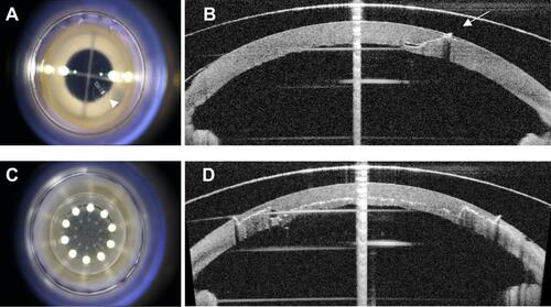
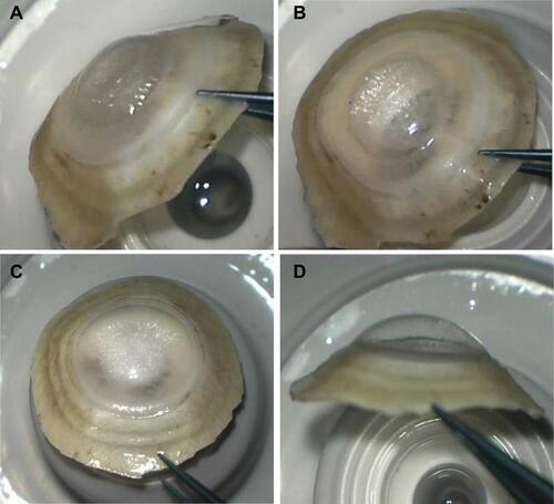
Figure 2 Appearance of type 1 and type 2 BB after eversion of the cornea. Type 1 BB, frontal (A) and lateral view (B); type 2 BB, frontal (C) and lateral view (D) showing a smaller diameter type 1 compared to type 2 bubble.