Figures & data
Figure 1 A schematic showing the theoretical mechanism of action. (A) posterior pole after staining, and (B) after ILM (internal limiting membrane)-rhexis, with vectors (arrows) of retinal relaxation after removal of ILM layer.

Table 1 Chronology of surgical procedures for each of the 14 cases
Table 2 Features of retinal detachment at time of surgery with ILM-peeling
Figure 2 ILM peeling over detached macula using asymmetric 25-gauge end-grasping forceps. View the surgical video using this link: http://youtu.be/7KWk2Jyngrs.
Abbreviation: ILM, internal limiting membrane.
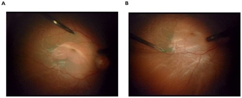
Table 3 Surgical features and clinical outcomes of study group