
Figures & data
Table 1 Baseline Characteristics of Study Participants
Figure 1 Kaplan–Meier survival curve of recurrence till the end of the follow-up period. Event refers to retinal re-detachment after silicone oil removal. The table below reports event-free survival after 2 and 3 years of follow-up.
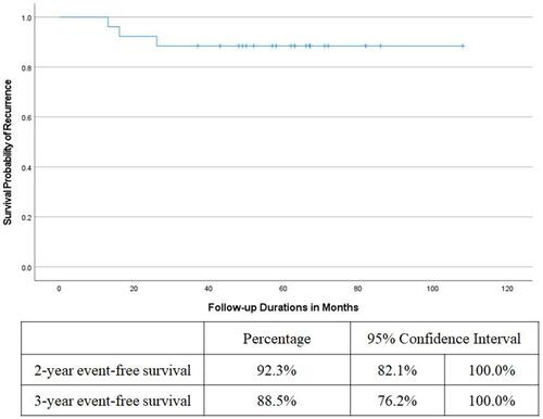
Figure 2 Left, pre-operative SD-OCT image in a 5-line raster scan mode of the right macula of a 62-year old female patient. Note the central retinal detachment and full-thickness macular hole. The axial length of the right eye measured 26.3 mm. Right, corresponding post-operative SD-OCT image in a 5-line raster scan mode. The patient had retinal re-attachment and U-type closure of the macular hole. Note the sub-foveal residual neuro-sensory retinal detachment and para-foveal cystoid changes. There is posterior bowing of the globe and significant thinning of the choriocapillaris, which are consistent with high myopia. Her final BCVA was 0.2 decimal units.
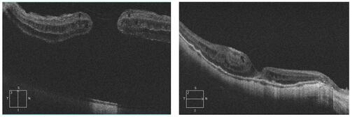
Figure 3 Post-operative SD-OCT image in a 5-line raster scan mode of the left macula of a 67-year old male patient. The patient presented with retinal detachment and macular hole. The axial length of the left eye measured 27 mm. Post-operatively he had retinal re-attachment and V-type closure of the macular hole. Note the dome-shaped macula, posterior bowing of the globe and significant thinning of the choriocapillaris, which are consistent with high myopia. His final BCVA was 0.05 decimal units.
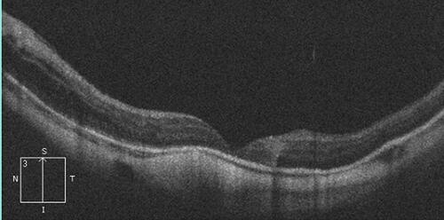
Figure 4 Post-operative SD-OCT image in a 5-line raster scan mode of the left macula of a 75-year old female patient. The patient presented with retinal detachment and macular hole. The axial length of the left eye measured 30 mm. Post-operatively she had retinal re-attachment and open-flat hole configuration (W-type). Note the characteristic steep concavity due to posterior bowing of the globe and significant thinning of the choriocapillaris, which are consistent with high myopia. Her final BCVA was 0.05 decimal units.
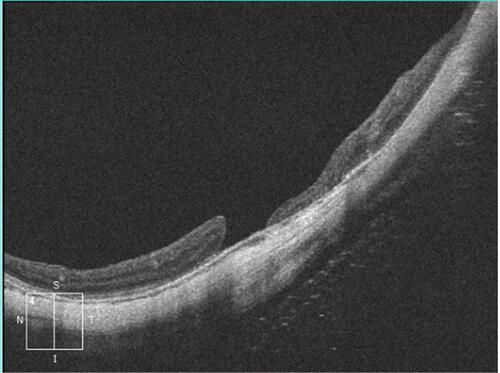
Table 2 Anatomical and Functional Post-Operative Outcomes
Table 3 Review of Studies on PPV and Silicone Oil Injection for High Myopic Retinal Detachment Associated with Macular Hole