Figures & data
Table 1 Patient Demographics, Characteristics and Aetiology of Neurotrophic Keratopathy (NK)
Figure 1 Preoperative neurotrophic keratopathy demonstrated by scarring and punctate keratopathy with improvement in corneal sensations and clear cornea in patient 6.
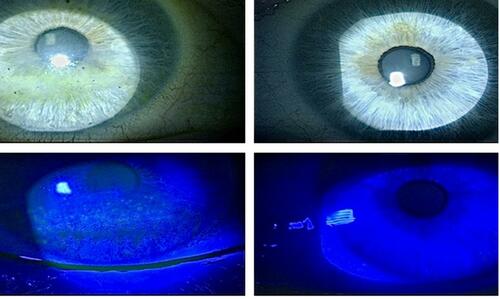
Table 2 Functional Outcome Data of Patients with Neurotrophic Keratopathy
Figure 2 In vivo confocal microscopy images of the subbasal nerve plexus for patient 7 taken at 50µm depth- Pre op presence of one main nerve fibre (arrow) (A); one main nerve fibre (arrow) and nerve branches (dotted arrow) at 3 month (B); one main nerve fibre (arrow) and multiple nerve branches (dotted arrow) at 6 month (C); three main nerve fibres and multiple nerve branches at 12 month (D) after corneal neurotisation surgery (-: main nerve, –: nerve branches showing nerve regeneration).
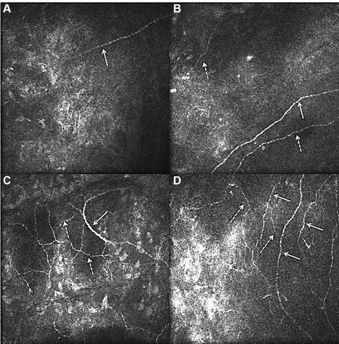
Table 3 Nerve Parameters of Five Patients with IVCM Data
Table 4 Nerve Parameters (Mean Change of Nerve Parameters) for the Five Patients at Different Postperative Time Points
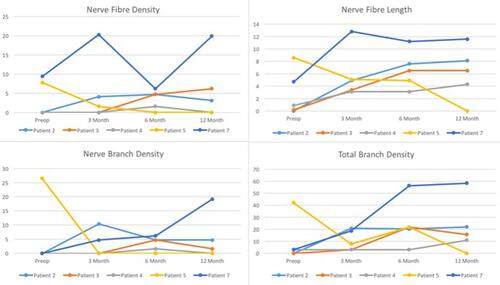
Figure 3 Nerve parameters (nerve fiber density, nerve fiber length, nerve branch density and total branch density) in patients 2, 3, 4, 5 and 7 improving from preoperative to early (3 month), intermediate (6 months) and late (12 months) postoperative follow-up.