Figures & data
Figure 1 Slit-lamp biomicroscopic findings in Case 1. (A) Conjunctival tumor can be seen at the corneal limbus with edema and feeder vessel (arrow). A petechial pigmented lesion can be seen (arrowheads). (B) Photograph of eye one month after treatment with topical epinastine and tacrolimus, an immunosuppressive drug. The tumor size and edema are markedly reduced, and the feeder vessel has regressed (arrow). (C) One year after the treatment. The tumor is not detectable.
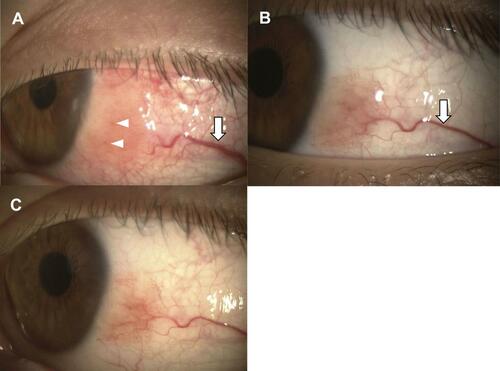
Figure 2 Slit-lamp biomicroscopic findings in Case 2. (A) Conjunctival tumor with high degree of pigmentation can be seen at the corneal limbus. There is also mild edema and feeder vessels (arrows). (B) The edema is reduced after one month of treatment with epinastine eye drops.
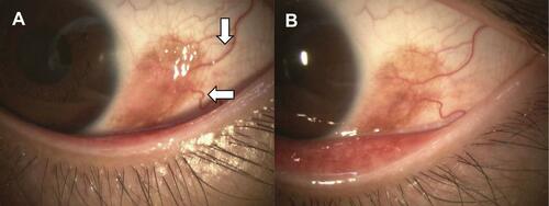
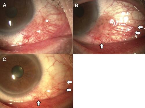
Figure 3 Slit-lamp biomicroscopic findings in Case 3. (A) There is marked hyperemia around the corneal limbus. Petechial pigmented lesion can be seen in the same area (arrow heads). (B) Photograph one month after treatment with topical epinastine and 0.1% fluorometholone eye drops. We can see an edematous neoplastic lesion with pigmentation, and it was accompanied by dilated feeder vessels (arrows). (C) One month after the treatment with topical epinastine and tacrolimus. The hyperemia, edema, and pigmentation are markedly improved and feeder vessels regressed (arrows).