Figures & data
Table 1 Tumor Details in OAL Patients
Table 2 Radiographic Findings in IgG4-ROD vs OAL Patients
Figure 1 Bilateral periocular tendon involvement in a patient with OAL presenting via blurry muscle insertions of the four recti muscles (coronary CT with contrast agent, soft tissue window).
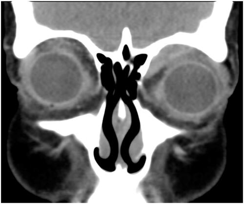
Figure 2 (A) Apparent bilateral infraorbital canal and nerve infiltration (arrows) in a patient with IgG4-ROD (coronary T1 MRI scan with contrast agent). (B) In comparison bilateral infraorbital nerve infiltration (arrows) in a patient with mantle cell OAL (coronary CT scan with contrast agent, soft tissue window).
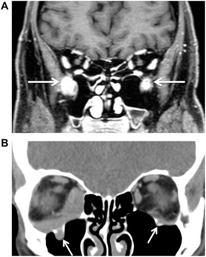
Figure 3 Bilateral infiltration of the orbital fat pad in a patient with OAL (axial CT scan with contrast agent, soft tissue window).
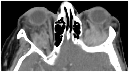
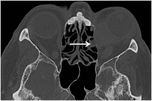
Figure 4 Infiltration of the lacrimal system and ethmoidal cells (arrow) in a patient with IgG4-ROD (axial CT scan with contrast agent, bone window).