Figures & data
Figure 1 83-year old female with exudative age-related macular degeneration of the right eye. (A) Fundus photograph of the left eye showed macular drusen at the time of presentation. (B) The right fundus photograph revealed large, thick submacular hemorrhage at the time of presentation (SMH). (C) OCT of the right eye from presentation revealed subretinal hemorrhage. (D)At one-month follow-up after receiving intravitreal bevacizumab, there was some resolution of the SMH with persistent dehemoglobinzied blood. (E) Six months later with continuation of anti-VEGF treatment, the hemorrhage had been completely resolved, with inferior subretinal fibrosis that was away from the fovea. (F) At ten-year follow-up, the inferior subretinal fibrosis developed pigmentation; (G) corresponding OCT was stable without subretinal fluid.
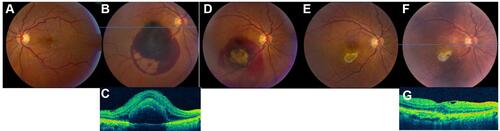
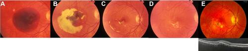
Figure 2 49-year old female with presumed ocular histoplasmosis syndrome (POHS) of the right eye. (A) On presentation, fundus photograph of the right eye revealed a large, thick SMH. There was no OCT imaging or anti-VEGF treatment agents available in 1989. (B) During one-month follow-up, while some of the hemorrhage had been resorbed, there was persistent dehemoglobinized blood. (C) At three-month follow-up, the hemorrhage had mostly resolved with minimal retinal pigment epithelial changes. (D) Two years later, hemorrhage was resolved completely with inferior subretinal fibrosis. (E) At 30-year follow-up, fundus photograph demonstrated pigmented fibrosis and the OCT showed a shallow RPE detachment without subretinal fluid.