Figures & data
Figure 1 Example of a print-out from the papillary anatomic and vascular assessments (RTVue XR Avanti, with AngioVue system, Optovue®).
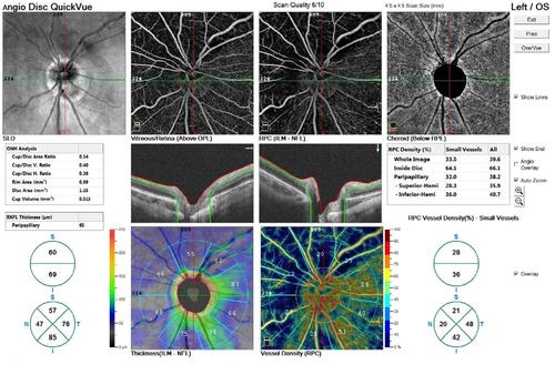
Figure 2 Example of a print-out from the circumpapillary microperimetric assessment (MP-3 microperimeter, Nidek®).
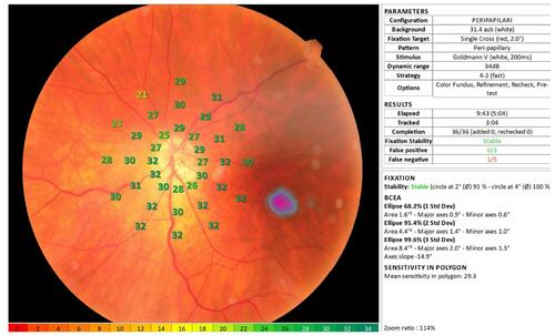
Table 1 Overall and per Group Analysis
Table 2 Discriminative Power of Each Variable
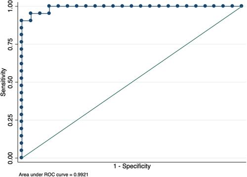
Figure 3 Best 2-variable papillary anatomic model to predict the presence of glaucoma in eyes with high myopia, including the peripapillary retinal nerve fiber layer thickness of the temporal and inferior sectors.
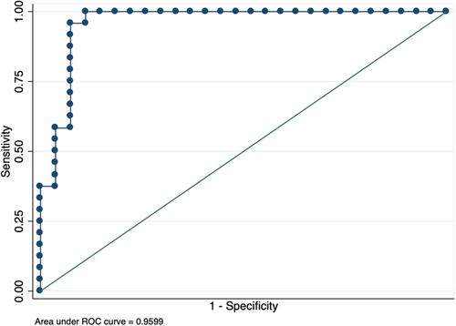
Figure 4 Best 2-variable papillary vessel density model to predict the presence of glaucoma in eyes with high myopia, including the en-face papillary retinal small vessel densities of the inferior and superior sectors.