Figures & data
Table 1 Demographic Data and Possible Predisposing Factors
Figure 1 Diagrams of infiltration pattern and clinical slit-lamp photographs. (A1 and A2) Diagram of a reticular infiltration pattern and a photograph of a 21-year-old student who had a history of contact-lens wearing and presented at KCMH two days after left eye irritation (Case#21); (B1 and B2) diagram of a satellite infiltration pattern and a photograph of a 55-year-old farmer who had a trauma to the right eye from grass leaves (Case#15); (C1 and C2) diagram of a multifocal infiltration pattern and a photograph of a 58-year-old farmer who had a trauma of the right eye with soil contamination (Case#24); (D1 and D2) diagram of a total infiltration pattern and a photograph of the right eye of a 61-year-old female without a significant history of ocular trauma (Case#10).
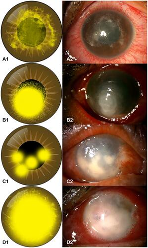
Table 2 Clinical and Diagnostic Data
Table 3 Demographic Data and histopathology of Globe Salvage Group versus Globe Removal Group (Categorical Variables)
Table 4 Demographic Data and Histopathology of Globe Salvage Group versus Globe Removal Group (Continuous Variables)
Figure 2 Box plot demonstrates that almost all of the cases from the globe salvage group received their first medical treatment on day 1 after onset of symptoms (asterisks indicate extreme outliers).
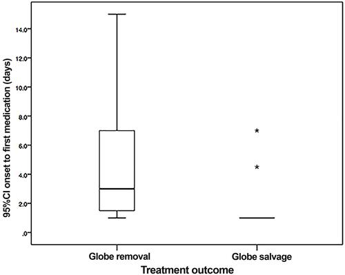
Figure 3 Histopathology of Pythium keratitis in a globe salvage case (Case#19). (A) Histopathology section shows an ulcerated corneal lesion with numerous acute inflammatory cells and necrotic cells primarily located at the anterior stroma (Hematoxylin-Eosin); (B) special stain shows varying sizes of short hyphae at the posterior stroma and pre-Descemet’s membrane area (Gomori Methenamine Silver).
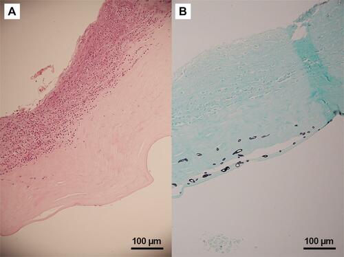
Figure 4 Histopathology of Pythium keratitis in globe salvage case (Case#05). (A) Histopathology section shows numerous acute inflammatory cells and necrotic cells in the corneal stroma (Hematoxylin-Eosin); (B) special stain shows varying-sized short hyphae at the mid stroma (Gomori Methenamine Silver).
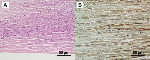
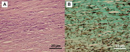
Figure 5 Histopathology of Pythium keratitis in globe removal case (Case#23). (A) Histopathology section shows numerous acute inflammatory cells and necrotic cells in the corneal stroma (Hematoxylin-Eosin); (B) special stain shows numerous varying-sized short hyphae with rare septation (Gomori Methenamine Silver).