Figures & data
Figure 1 The comparison of the retinal thickness in the fovea (RT1) and parafoveal region (RT2), as well as of the retinal nerve fiber layer thickness in the temporal, superior, nasal and inferior quadrant (RNFL-T, RNFL-S, RNFL-N, RNFL-I) between group S(-) and S(+).
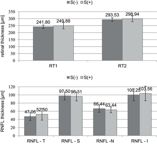
Figure 2 The comparison of mean PVEP P100-wave amplitudes (A) and latencies (L) at the 1°04ʹ and 0°16ʹ checkerboard check size between group S(-) and S(+).
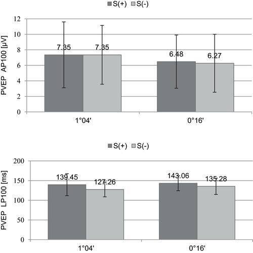
Figure 3 The comparison of mean PERG P50- and N95-wave amplitudes and P50-wave implicit times between group S(-) and S(+).
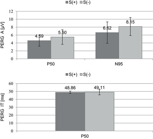
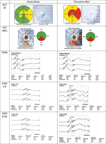
Figure 4 The results of macular (RT) and retinal nerve fiber layer (RNFL) thickness in optical coherence tomography (OCT), and pattern electroretinogram (PERG) and visual evoked potentials (PVEP) recordings from the patient’s left eye, with the history of not-treated demyelinating ON at the acute phase and 2 years later.