Figures & data
Table 1 Epidemiology of Microorganisms Found in Infectious Keratitis Cases
Figure 1 3 examples of green thermal laser application for resistant infectious keratitis (IK). Case 1 (A–C) had a bacterial IK, with a 1 mm height hypopyon level. Pre-treatment (A), post-laser (B), and after complete healing (C). Case 2 (D–F) had a fungal IK, with a 3 mm hypopyon level. Pre-treatment (D), immediately post-laser with stained cornea (E), and after complete healing (F), with residual corneal opacity. Case 3 (G–I) had mixed bacterial and fungal dense central infiltration with a flat anterior chamber (G), Post-laser (H), and after amniotic membrane transplantation (I).
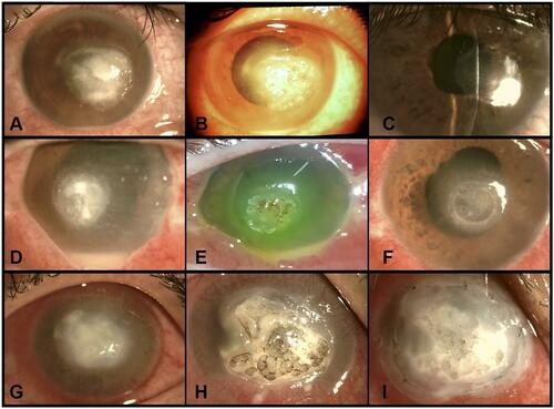
Figure 2 Duration till complete healing and full resolution of inflammatory signs after laser application, ranging between 2–6 weeks. The majority of cases had complete healing within the first 2 weeks (77 cases, 51.3%).
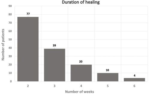
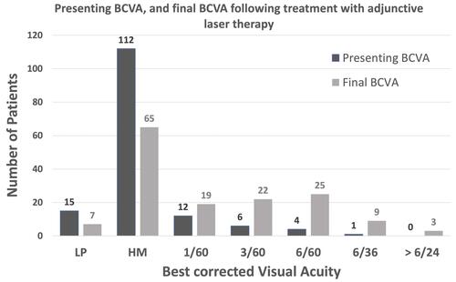
Figure 3 Presenting and final corrected distance visual acuity (CDVA) after adjunctive laser therapy. Before starting the treatment, 127 (84.6%) cases had a CDVA of hand movement (HM), or light perception (LP). After complete healing 78 (52%) of cases had a CDVA better than HM.