Figures & data
Figure 1 Images of macular region on spectral domain and swept source optical-coherence tomography (A, B, D: Spectralis OCT, Heidelberg Engeneering, Heidelberg, Germany; C: SS OCT; CArl Zeiss, Oberkochen, Germany).
Notes: (A) Attached posterior hyaloid. (B and C) Partial elevation of the nasal posterior vitreous (partial posterior vitreous detachment) can be seen as a thin layer (arrows) anterior of the retinal surface. Vitreous is still attached in foveal region. (D) Complete vitreous detachment, an entire separation between the posterior vitreous cortex (arrows) and the retina surface.
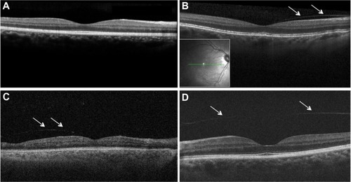
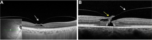
Figure 2 Images of macular region on spectral domain optical coherence tomography (SD-OCT; Spectralis OCT, Heidelberg Engeneering, Heidelberg, Germany).
Notes: (A) Vitreomacular traction with focal traction at foveal (arrow) region and intrafoveal pseudocysts. (B) Full-thickness macular hole with vitreomacular traction (yellow and white arrows, respectively).