Figures & data
Figure 1 Causes of corneal irregularity. n=57 eyes. 6 cases were binocular.

Figure 2 Safety. Changes in Snellen lines of CDVA at final follow-up (12 months). n=52.
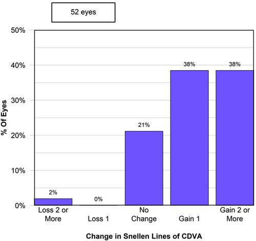
Figure 3 Improvement in CDVA (LogMAR) was significant at 3, and 6 months after surgery. CDVA stabilized after 6 months.
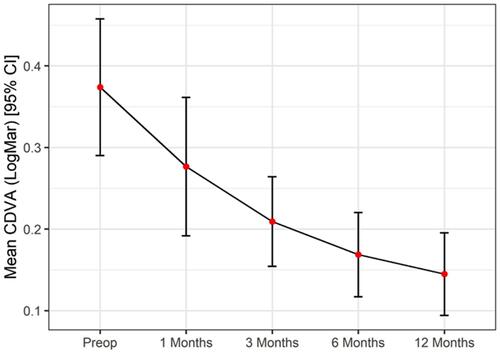
Figure 4 Kmean changes at 1, 3, 6, and 12 months. Changes proved to be significant up-to 3 months after surgery. Measurements performed with different devices are shown in different colors.
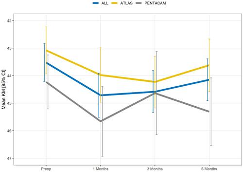
Figure 5 Mean corneal astigmatism measured with both Pentacam and Atlas 9000 topographers. Measurements performed with different devices are shown.
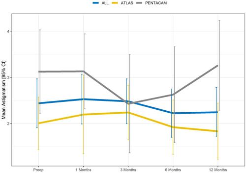
Table 1 Changes in Corneal Aberrations Measured with Both Devices. Parameters Estimated with Mixed Regression Model. Parameters are Given as Estimate (Standard Error)
Figure 6 Changes in corneal aberrations during the entire follow-up period. (A) No significant changes were observed in Total HOA. (B) Overall, changes in coma were significant at 6 months after surgery (P<0.01), and remained unchanged after. (C) No significant changes were observed in SA. Measurements performed with different devices are depicted.
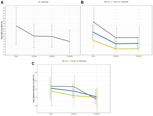
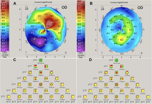
Figure 7 Preoperative and postoperative topographic, and aberrometric changes after TE-TG-PTK at 12 months follow-up. Sagittal maps (A, B) show substantial regularization of the corneal surface: Kmax decreased from 44.0 D preoperatively (A) to 41.5 D postoperative (B) without a major decrease in Kmean (41.3 D (A) to 40.8 D (B)). Zernike analysis (C, D) also shows considerable changes in total coma with figures decreasing from 2.84 um preoperatively (C) to 0.070 um (sum of the mean root square) at final follow-up (D). This patient had previous diagnosis of diffuse corneal leucomas secondary to adenoviral keratoconjunctivitis, and gained 6 lines of Snellen CDVA (from LogMAR 1.92 to −0.1) at final follow-up.