Figures & data
Figure 1 Representative photographs during the surgery and a schematic illustration of each incision type used; (A) a conventional 5.5-mm sclerocorneal incision, (B) L-shaped incision, and (C) frown incision.
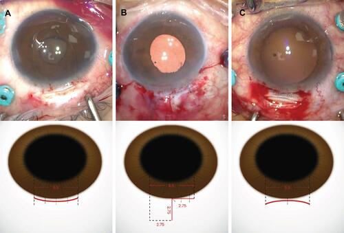
Table 1 Demographic and Preoperative Ocular Characteristics of Enrolled Patients (n=107)
Figure 2 Standard graphs for reporting refractive outcomes. (A) Uncorrected distance visual acuity (UDVA). (B) The difference between UDVA and corrected distance visual acuity (CDVA). (C) Change in CDVA, (D) The comparison between attempted spherical equivalent (SEQ) refraction and achieved SEQ refraction. (E) The accuracy of SEQ to the intended target. (F) Spherical equivalent refraction stability after surgery. (G) The comparison between preoperative and postoperative refractive astigmatism.
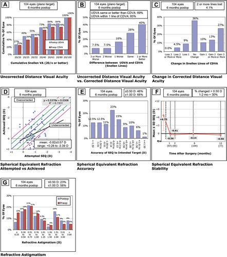
Table 2 Comparison of the Postoperative 6 Month Analysis (n=107)
Figure 3 Cumulative histogram of the magnitude of the preoperative corneal and postoperative refractive astigmatism, vertexed to the corneal plane; (A) a conventional 5.5-mm sclerocorneal incision, (B) L-shaped incision, and (C) frown incision.
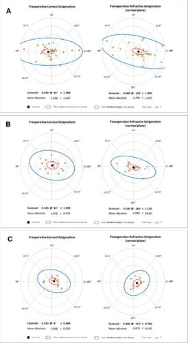
Table 3 Comparisons of the Postoperative Complications (n=107)
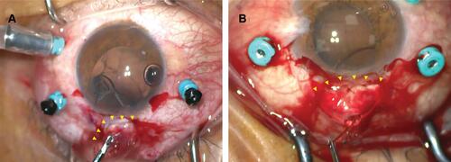
Figure 4 Representative surgical images of the (A) L-shaped incision and (B) frown incision.