Figures & data
Figure 1 Large area of implant exposure: implant removal and DFG is advisable. Note: Image is the property of the authors.
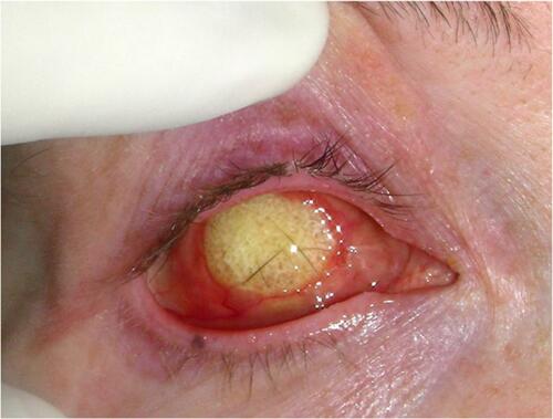
Figure 2 (A) Left orbital volume deficiency post left enucleation with no primary implant. (B) Adequate orbital volume following left secondary ball implantation. (Image is the property of the authors).
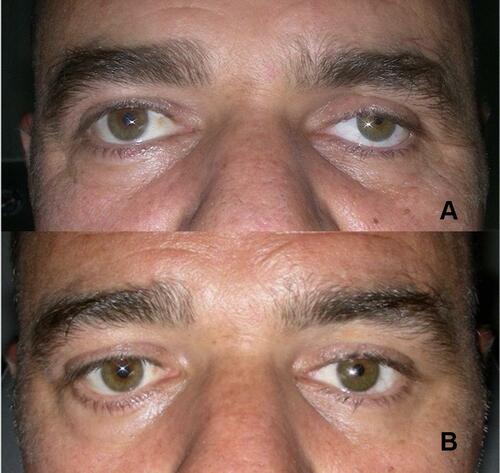
Figure 3 Bare DFG positioned in the socket with slight volume oversizing. (Image is the property of the authors).
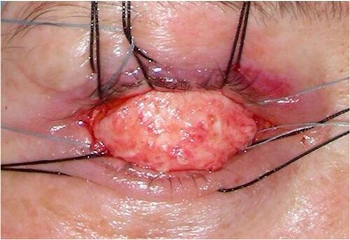
Figure 4 DFG to upper sulcus. (Image is the property of the authors).
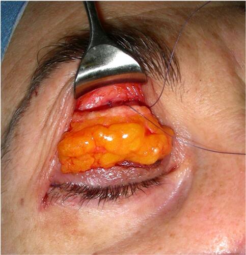
Figure 5 System for socket volume enhancement. (Image is the property of the authors).
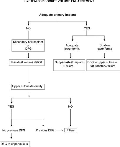
Figure 6 (A) Right ptosis and lower lid laxity following secondary implantation. (B) Ptosis corrected simultaneously with right and left lower lid malpositions. (Image is the property of the authors.
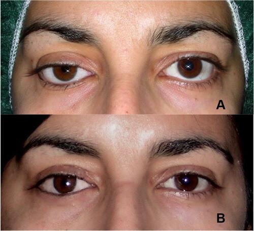
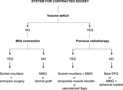
Figure 7 System for contracted socket. (Image is the property of the authors).