101
Views
3
CrossRef citations to date
0
Altmetric
Original Research

Visual Outcomes and Prognostic Factors of Large Submacular Hemorrhages Secondary to Polypoidal Choroidal Vasculopathy

, ORCID Icon, , , , , & show all
Pages 3557-3562 | Published online: 24 Aug 2021

Figures & data

Figure 1 A representative case of a large submacular hemorrhage (SMH) secondary to polypoidal choroidal vasculopathy. (A) A color fundus photograph shows a large SMH. (B and C) Infrared reflectance and optical coherence tomography (OCT) images show the SMH and hemorrhagic retinal pigment epithelial detachment. (B) The green arrow indicates the direction of OCT scan in (C). (D and E) Fluorescein and indocyanine green angiography performed after pneumatic displacement. (E) The yellow arrow indicates a polypoidal lesion. (F and G) Fluorescein angiography and OCT images performed after pneumatic displacement. (F) The green arrow indicates the direction of the OCT scan in (G). (G) The yellow arrow indicates the polypoidal lesion.

Figure 1 A representative case of a large submacular hemorrhage (SMH) secondary to polypoidal choroidal vasculopathy. (A) A color fundus photograph shows a large SMH. (B and C) Infrared reflectance and optical coherence tomography (OCT) images show the SMH and hemorrhagic retinal pigment epithelial detachment. (B) The green arrow indicates the direction of OCT scan in (C). (D and E) Fluorescein and indocyanine green angiography performed after pneumatic displacement. (E) The yellow arrow indicates a polypoidal lesion. (F and G) Fluorescein angiography and OCT images performed after pneumatic displacement. (F) The green arrow indicates the direction of the OCT scan in (G). (G) The yellow arrow indicates the polypoidal lesion.

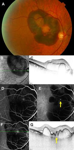
Table 1 Characteristics of Patients with Large SMHs Secondary to PCV

Figure 2 The best-corrected visual acuities (BCVAs) expressed in logarithm of the minimum angle of resolution (logMAR) units (mean ± standard deviation) at the development, 1 month, and 1 year after the development of large submacular hemorrhages, and at the final visit.

Figure 2 The best-corrected visual acuities (BCVAs) expressed in logarithm of the minimum angle of resolution (logMAR) units (mean ± standard deviation) at the development, 1 month, and 1 year after the development of large submacular hemorrhages, and at the final visit.

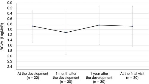
Table 2 Prognostic Factors Predictive of BCVA 1 Year After Development of Large SMHs Secondary to PCV