Figures & data
Table 1 Equipment Settings
Table 2 NGENUITY 3D DAVS Settings Applied During Surgery
Figure 1 Residual vitreous assessment. Posterior segment of the eye was removed (A), the eye was bisected (B), and residual vitreous was stripped (C and D).
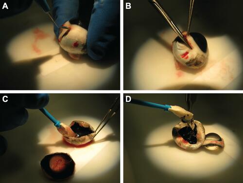
Figure 2 Comparison of NGENUITY 3D DAVS and a standard visualization system for the removal of vitreous. Mean residual vitreous (A) and mean percentage of vitreous removed (B) are shown (n = 15). *P < 0.0001.
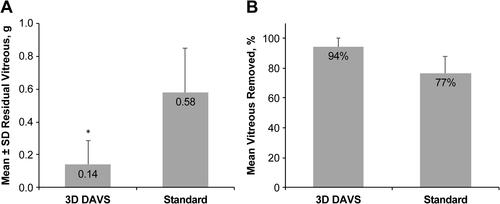
Table 3 Comparison of OD and OS
Table 4 Vitreous Weight and Axial Length for Contralateral Eyes in Comparison Analysis
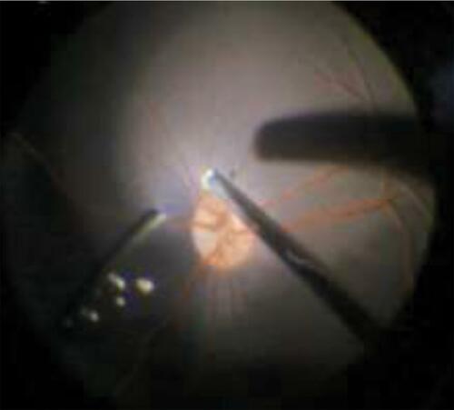
Figure 3 Vitreous visualization using NGENUITY 3D digitally assisted visualization system alternated between right eye and left eye to reduce surgical bias; vitreous was highlighted in a blue color.