
Figures & data
Figure 1 Diagram detailing the experimental steps for the elimination of bacteria attached to corneoscleral tissue by topical antiseptics.
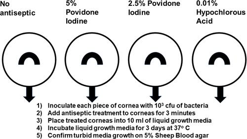
Table 1 Comparison of 5% and 2.5% Povidone Iodine Antibacterial Efficacy to 0.01% Hypochlorous Acid Using Bacteria Isolated from Endophthalmitis. Corneoscleral Tissue Was Used as a Solid-Phase Medium to Assimilate the Ocular Surface. Prevention of Bacterial Growth After Antiseptic Application Indicated Success of the Antiseptic
Table 2 Weighted Comparison of 5% and 2.5% Povidone Iodine Antibacterial Activity to 0.01% Hypochlorous Acid Based on Bacteria Isolated from Endophthalmitis Over a 27-Year Period (1993–2019). Corneoscleral Tissue Was Used as a Solid-Phase Medium to Assimilate the Ocular Surface. Prevention of Bacterial Growth After Antiseptic Application Indicated Success of the Antiseptic