Figures & data
Figure 1 Schematic representation of melanin biosynthesis in the melanocyte. After synthesis and subsequent processing in the Golgi body - rough endoplasmic reticulum (ER) complex, tyrosinase and tyrosinase-related protein 1 (TYRP1) are trafficked to the developing melanosome via membrane transporter proteins P and MATP. Mutations in tyrosinase or in TYRP1 result in the retention of these mutant proteins in the ER, and these are subsequently degraded using proteasome mediated pathways. The rest of the tyrosinase and TYRP 1 proteins undergo endosomal/lysosomal proteolysis. Retinal pigment epithelium makes and releases L-DOPA during the procerss of melanin biosynthesis; L-DOPA is an endo agonist for GPR 143 (expressed at the membrane of the melanosomes) thus create an autocrine loop. GPR143 is the protein product of OA1 and mutations at this locus result in ocular albinism type 1.
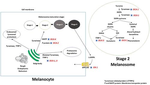
Table 1 Summary of Genetic, Pathophysiologic and Clinical Features Associated with Syndromic OCA
Figure 2 Iris transillumination defect, albinotic fundus and foveal hypoplasia in pseudophakic eyes of a 41-year-old patient. Note that major retinal blood vessels show a relatively wide exit angle as they emerge from the optic disc.
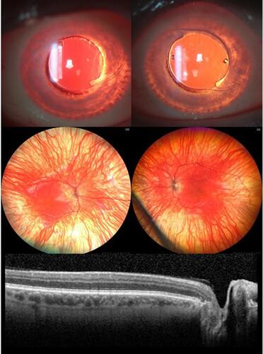
Figure 3 Schematic of normal and albino visual pathways. (A) Normal visual pathways. Nerve fibres originating from temporal retina project to the ipsilateral hemisphere (red line). Nerve fibres originating from predominantly nasal retina, cross at the chiasm and project to the contralateral hemisphere (blue line). (B) Albino misrouting. The majority of optic nerve fibres decussate to the contralateral hemisphere (red and blue lines). Adapted from Neveu MM, Jeffery G. Chiasm formation in man is fundamentally different from that in the mouse. Eye. 2007;21(10):1264–1270.Citation86
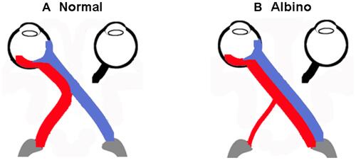
Figure 4 Normal Flash VEPs (A) P2 peak time in ipsilateral and contralateral hemispheres of control subjects. Peak times from both hemispheres are symmetrical and shorten until approximately 18–20 years of age, do not change between 20–40 years of age, then increases beyond 40 years of age (B) P2 amplitude in ipsilateral and contralateral hemispheres of control subjects. P2 amplitudes from both hemispheres are symmetrical and reduce significantly with age (P < 0.05). Reprinted from Neveu MM, Jeffery G, Burton LC, Sloper JJ, Holder GE. Age-related changes in the dynamics of human albino visual pathways. Eur J Neurosci. 2003;18(7):1939–1949.Citation110
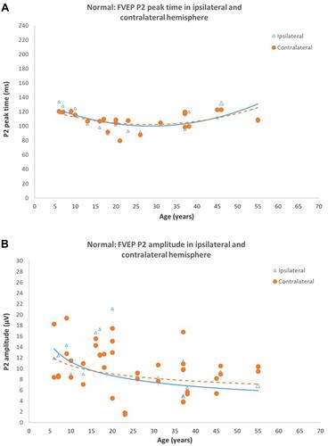
Figure 5 Normal Pattern onset-offset VEPs (A) CII peak times in ipsilateral and contralateral hemispheres of control subjects. CII peak times from both hemispheres are symmetrical and significantly shorten with increasing age (P < 0.0001). (B) CII amplitude in ipsilateral and contralateral hemispheres of control subjects. CII amplitudes from both hemispheres are symmetrical. P2 amplitude reduces significantly with age (P < 0.005). Reprinted from Neveu MM, Jeffery G, Burton LC, Sloper JJ, Holder GE. Age-related changes in the dynamics of human albino visual pathways. Eur J Neurosci. 2003;18(7):1939–1949.Citation110
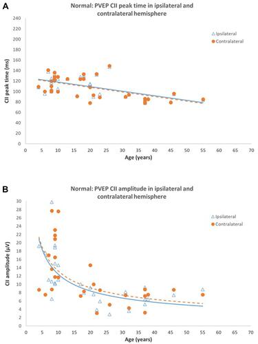
Figure 6 Pattern onset-offset VEPs and Flash VEPs from an age-matched control (A) and a 60 year old patient with albinism (B, C) using five scalp electrodes positioned over the occiput; 2 electrodes over the right hemisphere; two electrodes over the left hemisphere; 1 electrode over the midline. (A) VEP responses from control subject are symmetrical and of similar amplitude and peak time from the left and right hemisphere. (B). Pattern onset-offset VEP responses from a 60-year-old patient with albinism. Right eye (rows 2) VEP responses are of shorter peak and/or larger amplitude from the left hemisphere compared with the right hemisphere. Left eye (rows 3) VEP responses are of shorter peak and/or larger amplitude from the right hemisphere compared with the left hemisphere. (C) Flash VEP responses from a 60-year-old patient with albinism. VEP responses from both eyes are symmetrical and of similar amplitude and peak time from the left and right hemisphere.

Figure 7 Flash VEPs from an 8-month-old patient with albinism using five occipital scalp electrodes; 2 electrodes over the right hemisphere; 2 electrodes over the left hemisphere; 1 electrode over the midline. Right eye (row 1) VEP responses are of higher amplitude from the left hemisphere compared with the right hemisphere. Left eye (row 2) VEP responses are of higher amplitude from the right hemisphere compared with the left hemisphere.
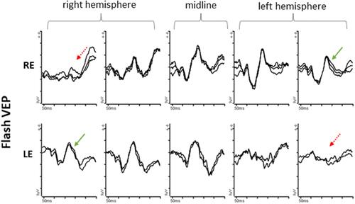
Figure 8 Flash VEP characteristics in 83 patients with albinism including 17 genetically confirmed cases of OCA, 3 cases of OA, 5 cases of HP syndrome and 1 case of CH Syndrome. (A) P2 amplitude in ipsilateral (bold line) and contralateral (dashed line) hemispheres. There is a significant inter-hemispheric amplitude difference up until ~18 years of age. There is no significant inter-hemispheric difference beyond 18 years. (B) Flash VEP P2 peak time in ipsilateral (bold line) and contralateral (dashed line) hemispheres of patients with albinism. P2 peak times in both hemispheres are symmetrical and similar to those in control subjects. Black filled data points are from genetically confirmed patients. Data from Neveu MM, Jeffery G, Burton LC, Sloper JJ, Holder GE. Age-related changes in the dynamics of human albino visual pathways. Eur J Neurosci. 2003;18(7):1939-1949Citation110, with additional patients and highlighting of genetically confirmed cases.
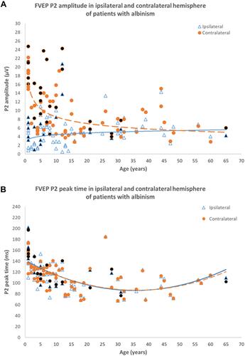
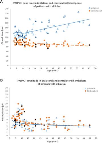
Figure 9 Pattern onset-offset VEP characteristics in 66 patients with albinism including 8 genetically confirmed cases of OCA, 3 cases of OA, 2 cases of HP syndrome and 1 case of CH Syndrome. (A) CII peak time in ipsilateral (bold line) and contralateral hemispheres (dashed line). CII peak time in the contralateral hemisphere is shorter than peak time in the ipsilateral hemisphere for all ages. The inter-hemispheric peak time difference increases with age. (B) CII amplitude in ipsilateral (bold line) and contralateral (dashed line) hemispheres of patient with albinism. CII amplitudes in both hemispheres are symmetrical, similar to that in control subjects. Black filled data points are from genetically confirmed patients. Data from Neveu MM, Jeffery G, Burton LC, Sloper JJ, Holder GE. Age-related changes in the dynamics of human albino visual pathways. Eur J Neurosci. 2003 Oct;18(7):1939-1949Citation110, with additional patients and highlighting of genetically confirmed cases.