Figures & data
Table 1 Common Clinical Presentation of Lacrimal Gland Lesions
Table 2 Histopathological Distribution of Lacrimal Gland Lesions
Figure 1 (A) Histopathology of Lacrimal Gland showing perivascular granulomatous inflammation with eosinophilic infiltration in Eosinophilic granulomatosis with polyangiitis (Haematoxylin-Eosin stain (H&E), magnification X 200) (B) Angiolymphoid Hyperplasia with Eosinophilia showing endothelial proliferation with scattered eosinophils H&E, magnification X 200 (C) Cluster of Differentiation. CD34 stain showing endothelial proliferation with plumped endothelial cells of the same patient.
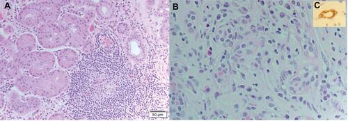
Figure 2 Pleomorphic Adenoma of The Lacrimal Gland; (A) Coronal fat saturated T2-WI and Apparent Diffusion Weight (ADC) image with lobulated outline, (B) Non-restricted pattern on Diffusion Weight Image (DWI).
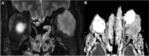
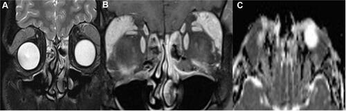
Figure 3 Right lacrimal gland enlargement with subtle enlargement of the right medial rectus muscle sparing the tendinous insertion in Thyroid Related Orbitopathy (TRO).

Figure 4 Chronic inflammation showing mixed lymphocytes and plasma cells infiltration (Haematoxylin-Eosin Stain H&E, magnification X100).
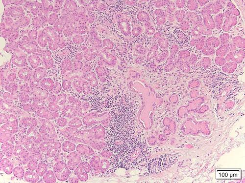
Figure 5 MRI of Extranodal Marginal Zone Lymphoma (A) Coronal fat saturated and post contrast (B) Fat suppressed T1 with contrast and apparent diffusion coefficient images (C) Diffusion Weight Image showing restricted pattern (mean 0.85 X 10_3).