Figures & data
Table 1 Patient Demographics and Previous Ocular History
Table 2 Retinal Pathology for Surgical Indication of Vitrectomy
Table 3 Quality of OCT Results Among Study Participants
Table 4 Status of Posterior Hyaloid After TA Staining
Table 5 Overall Sensitivity and Specificity of Optical Coherence Tomography versus Triamcinolone Staining
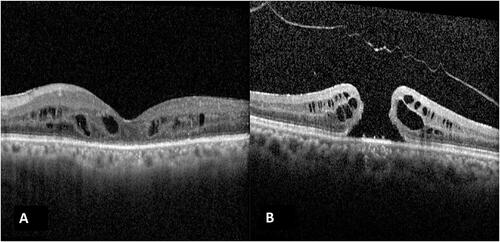
Figure 1 OCT image of a case labelled as Grade 4 PVD on OCT (A), but intraoperative TA staining determined a posterior hyaloid attachment (false-positive). Case (B) was labelled to have Grade 3 OCT (attached to the optic nerve head) although intraoperative staining showed that a complete PVD was present (false-negative).